

Perez-Almodovar, Tao, and Carta have observed the progress of the adsorption on a monoclonal antibody (mAb) from
Question:
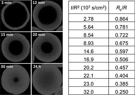
Perez-Almodovar, Tao, and Carta have observed the progress of the adsorption on a monoclonal antibody (mAb) from an aqueous solution at 25°C into spherical adsorbent particles using confocal laser scanning microscopy (CLSM). CLSM allows us to image optical sections revealing the distribution of the bound protein at the particle equator as a function of time. Some of the experimental results are shown in Fig. P.11.9. In these experiments, the mAb concentration in solution was kept constant at 1.0 mg/cm3 and the binding capacity, determined from independent measurements, was 110 mg/cm3.
The particles were around 54 μm in radius. Note that time is normalized by the square of the particle radius in order to account for some small differences between the different particles imaged.
(a) Using these data, plot the dimensionless position of the adsorption front ![]() as a function of
as a function of ![]() .
.
(b) As suggested by the analysis conducted in Problem 11.8, replot the data by graphing the function ![]() vs. the quantity
vs. the quantity ![]() and determine the effective pore De diffusivity of the mAb in these particles from the slope of this plot.
and determine the effective pore De diffusivity of the mAb in these particles from the slope of this plot.
(c) Assuming that the particles have a porosity ![]() and a pore radius of 50 nm, determine the tortuosity factor
and a pore radius of 50 nm, determine the tortuosity factor ![]() for these particles.
for these particles.
FIGURE P.11.9:
Step by Step Answer:

This question has not been answered yet.
You can Ask your question!


Heat And Mass Transfer For Chemical Engineers Principles And Applications
ISBN: 9781264266678
1st Edition
Authors: Giorgio Carta
