

Most humans are infected with herpes simplex virus-1 (HSV-1), the causative agent of cold sores. The HSV-1
Question:
Interestingly, only a single viral transcript is expressed during latency. This transcript, LAT (latency-associated transcript), does not encode a protein, and neurons infected with mutant HSV - I lacking the LA T gene undergo cell death by apoprosis at a rate twice that of cells infected with wild type HSV-1. To determine if LAT functions to block apoptosis by encoding a miRKA, the following studies were done (see Gupta et al., 2006, Nature 442:82-85).
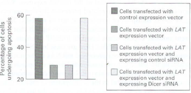
a. A cell line was transfected (a process in which foreign DNA is inserted into a cell) with an expression vector that expresses a Pst-Miu fragment of the LAT gene (see diagram in part b). The percentage of these transfected cells that then underwent drug-induced cell death was compared to that of control cells. The experiment was repeated in cells in which Dicer expression was knocked down using Dicer siRNA. The data obtained are shown in the graph below. What conclusions can be drawn from these data? Why did the scientists who conducted this study examine the effects of silencing Dicer?
b. Cells were transfected with an expression vector expressing the Pst-Miu fragment of the LAT gene from which the region between the two Sty restriction sites was deleted (DSty; diagram below). When these cells were induced to undergo apoptosis, they died at the same rate as did non-transfected cells. In additional studies, cells were transfected with an expression vector expressing the Sty-Sty region of the LA T gene. These cells exhibited the same resistance to apoptosis as did cells transfected with the Pst-Mlu fragment. What can be deduced from these findings about the region of the LAT gene required to protect cells from apoptosis?
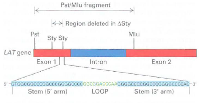
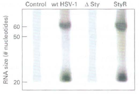
c. RNA encoded within the Sty-Sty region is predicted to form a stem loop (see diagram in part b). Northern blot analysis was performed on total-cell RNA isolated from control cells (mock), cells infected with wild-type HSV-1, cells infected with an HSV-1 deletion mutant from which the sequence between the two Sty site:, in the LA T gene was deleted (DSty), and cells infected with a rescued DSry virus into which the deleted region was re-inserted into the viral genome (StyR). The probe used for the Northern blot was the labeled 3' stem region of the LAT RNA in the Sty-Sty region, as diagrammed in part (b). The RNAs recognized by this probe were either ˆ¼ 5 nucleotides or 20 nucleotides, as shown in the Northern blot below. Why were two differentized RNAs detected? When a second probe was used that was the labeled 5' stem region of the RNA sequence shown in part (b), only the -55-nucleotide RNA was detected. What can you deduce about the processing of RNA expressed from the LAT gene? What enzyme likely produced the -55-nucleotide RNA? In what part of the cell? What enzyme likely produced the 20- nucleotide RNA? In what part of the cell?
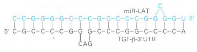
d. T GF-13 mRNA encodes a protein, transforming growth factor 13, that inhibits cell growth and induces apoptosis. The 3' untranslated region (3' UTR) of TGF-13 mRNA can form an imperfect duplex with miRNA encoded by the 5' stem region of the LA T Sty-Sty domain (miR-LAT), as shown below. In what way might the expression levels of TGF-13 differ in cells infected with wild-type HSV-1 compared to uninfected cells? What can you infer about latent HSV-1 infections from these studies?
Step by Step Answer:
a Dicer is needed to cleave microRNAs from a larger transcript The scientists tested the hypothesis that LAT encodes an miRNA by interfering with Dicer expression Without Dicer there should be no miRN...View the full answer


Molecular Cell Biology
ISBN: 978-1429234139
7th edition
Authors: Harvey Lodish, Arnold Berk, Chris A. Kaiser, Monty Krieger, Anthony Bretscher, Hidde Ploegh, Angelika Amon, Matthew P. Scott
