

The behavior of receptor X (XR), a transmembrane protein present in the plasma membrane of mammalian cells,
Question:
a. Cells expressing GFP-XR or artificial lipid vesicles (liposomes) containing GFP-XR are subjected to fluorescence recovery after photobleaching (FRAP). The intensity of the fluorescence of a small spot on the surface of the cells (solid line) or on the surface of the liposomes (dashed line) is measured prior to and following laser bleaching (arrow). The data are shown below.
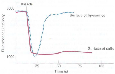
What explanation could account for the differing behavior of GFP-XR in liposomes versus in the plasma membrane of a cell?
b. Tiny gold particles can be attached to individual molecules and their movement then followed in a light microscope by single-particle tracking. This method allows one to observe the behavior of individual proteins in a membrane. The tracks generated during a 5-second observational period by a gold particle attached to XR present in a cell (left) or in a liposome (middle) or to XR adhered to a microscope slide (right) are shown below.
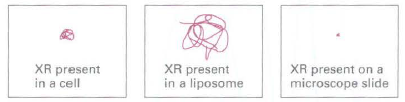
What additional information do these data provide beyond what can be determined from the FRAP data?
c. Fluorescence resonance energy transfer (FRET) is a technique by which a fluorescent molecule, following its excitation with the appropriate wavelength of light, can transfer its emission energy to and excite a nearby different fluorescent molecule. Cyan fluorescent protein (CFP) and yellow fluorescent protein (YFP) are related to GFP but fluoresce at cyan and yellow wavelengths rather than at green. If CFP is excited with the appropriate wavelength of light and a YFP molecule is very near, then energy can be transferred from CFP emission and used to excite YFP, as indicated by a loss of emission of cyan fluorescence and an increase in emission of yellow fluorescence.
CFP-XR and YFP-XR are expressed together in a cell line or are both incorporated into liposomes. The number of molecules of YFP-XR and CFP-XR per cm2 of membrane is equivalent in the cells and the liposomes. The cells and liposomes are then irradiated with a wavelength of light that causes CFP but not YFP to fluoresce. The amount of cyan (CFP) and yellow (YFP) fluorescence emitted by the cells (solid line) or liposomes (dashed line) is then monitored, as shown below.
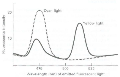
What can be deduced about XR from these data?
Step by Step Answer:
a In the liposomes there are no constraints on the diffusion of GFPXR in the plane of the bilayer Ac...View the full answer


Molecular Cell Biology
ISBN: 978-1429234139
7th edition
Authors: Harvey Lodish, Arnold Berk, Chris A. Kaiser, Monty Krieger, Anthony Bretscher, Hidde Ploegh, Angelika Amon, Matthew P. Scott
