

Question: 1. (3 points) For a conventional prostate cancer detection, a pathologist needs to inspect a digitized microscopy image of a prostate tissue slide (a sample
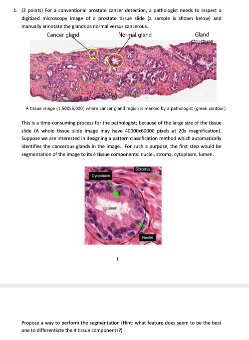
1. (3 points) For a conventional prostate cancer detection, a pathologist needs to inspect a digitized microscopy image of a prostate tissue slide (a sample is shown below) and manually annotate the glands as normal versus cancerous. Cancer gland Normal gland Gland structure A tissue image (1,500x5,000) where cancer gland region is marked by a pathologist (green contour) This is a time-consuming process for the pathologist; because of the large size of the tissue slide (A whole tissue slide image may have 40000x60000 pixels at 20x magnification). Suppose we are interested in designing a pattern classification method which automatically identifies the cancerous glands in the image. For such a purpose, the first step would be segmentation of the image to its 4 tissue components: nuclei, stroma, cytoplasm, lumen. Stroma Cytoplasm Lumen Nuclei 1 Propose a way to perform the segmentation (Hint: what feature does seem to be the best one to differentiate the 4 tissue components?)
Step by Step Solution
There are 3 Steps involved in it

Get step-by-step solutions from verified subject matter experts