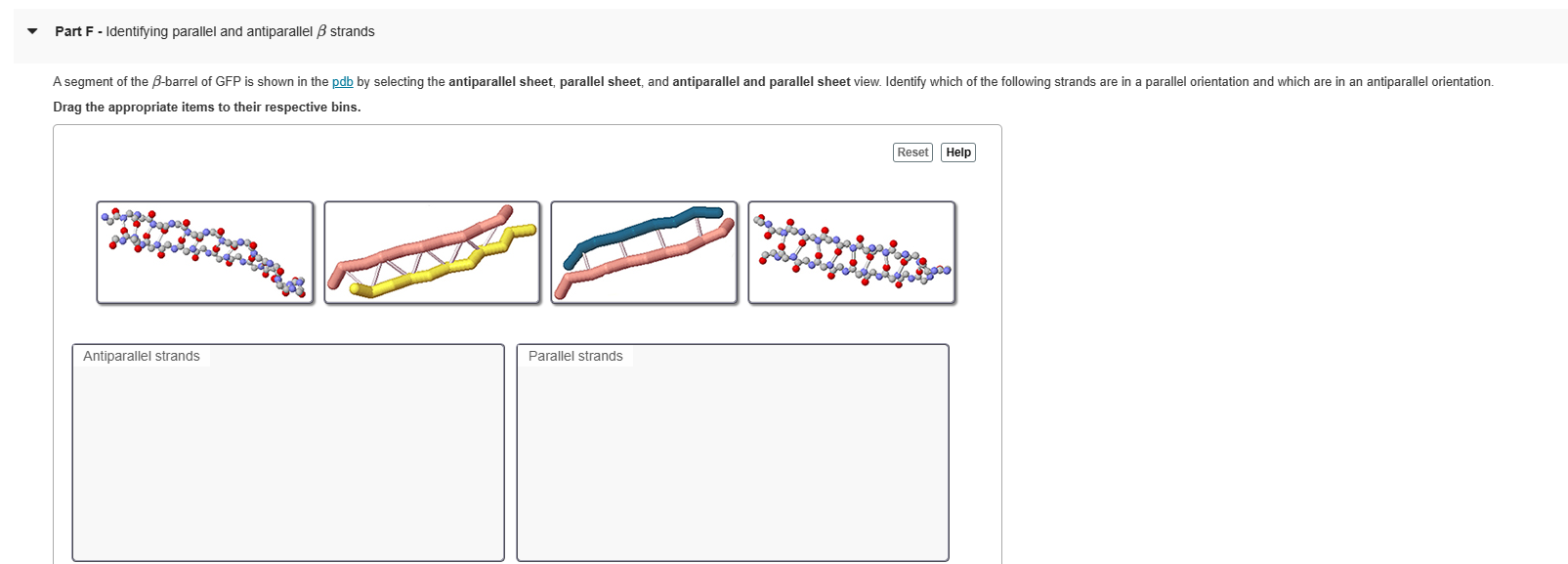
Question: Part F - Identifying parallel and antiparallel B strands A segment of the B-barrel of GFP is shown in the pdb by selecting the antiparallel

Part F - Identifying parallel and antiparallel BPart F - Identifying parallel and antiparallel BPart F - Identifying parallel and antiparallel B
Part F - Identifying parallel and antiparallel B strands A segment of the B-barrel of GFP is shown in the pdb by selecting the antiparallel sheet, parallel sheet, and antiparallel and parallel sheet view. Identify which of the following strands are in a parallel orientation and which are in an antiparallel orientation. Drag the appropriate items to their respective bins. Reset Help Antiparallel strands Parallel strandsLoops and turns Loops and turns are peptide sequences that join helices and sheets. Turns are typically short peptide segments that remain rigid in the structure. B turns join two B strands aligned in an antiparallel orientation. Helical bundles are often joined by short turns. Often hydrogen bonds hold the turn in a rigid position. Loops, in contrast, are typically longer, are located on the surface of the structure, and contain predominantly hydrophobic residues. They are flexible, not tied into the structure with lots of hydrogen bonding, and frequently are involved in the working end of proteins, changing shape in the presence or absence of a substrate or other ligand. You'll encounter a number of activation loops as you explore proteins. Part H - Distinguishing loops from turns Let's explore the structure of malate dehydrogenase. After opening the pdb, select the view secondary structure to view the carbon backbone. helix hydrogen bond loop/turn After analyzing the structure, predict which of the highlighted sections from the view highlighted loops and turns are loops or turns. Drag the appropriate items to their respective bins. Reset Help Most likely a loop Most likely a turn Uncertain classificationPart C - Exploring antiparallel B sheet structure The hydrogen bonding between antiparallel sheets, also visible in the pdb by selecting the antiparallel sheet view, is shown. Locate the N- and C-termini in this image. Select the antiparallel sheet view in the pdb file. How many peptide chains are shown? O 1 O 2 O 3 0 4

Step by Step Solution

There are 3 Steps involved in it

1 Expert Approved Answer
Step: 1 Unlock blur-text-image
Question Has Been Solved by an Expert!

Get step-by-step solutions from verified subject matter experts

Step: 2 Unlock
Step: 3 Unlock

Students Have Also Explored These Related Law Questions!