

Question: please help with this question with detailed solution thanks Question 9 We have developed an optical imaging system for single-cell inspection. Each cell can be
please help with this question with detailed solution thanks
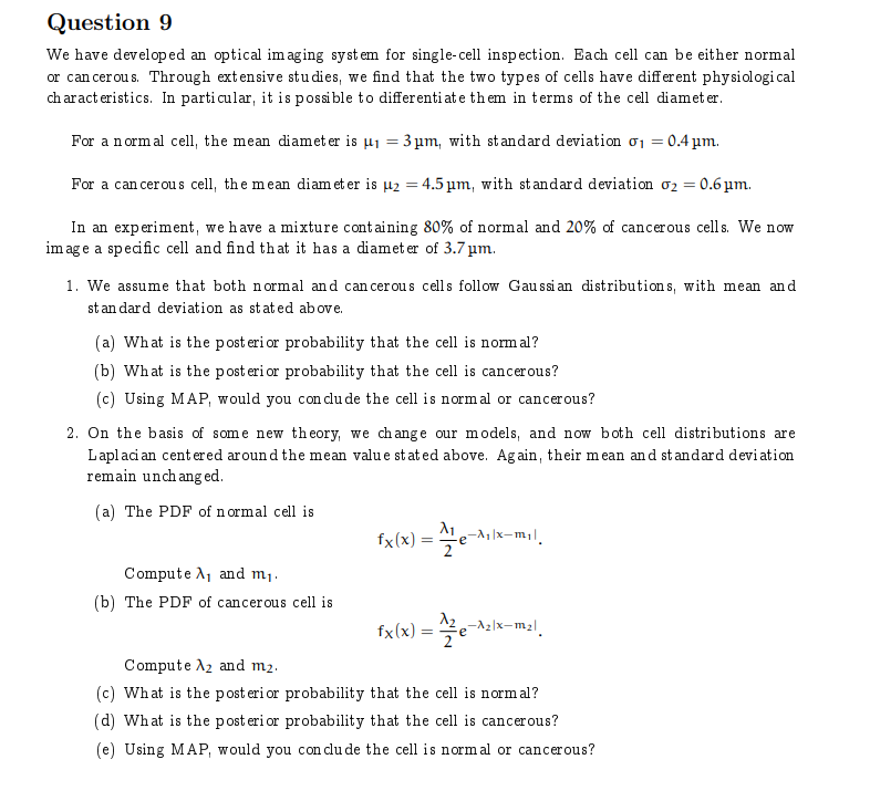
Question 9 We have developed an optical imaging system for single-cell inspection. Each cell can be either normal or cancerous. Through extensive studies, we find that the two types of cells have different physiological characteristics. In particular, it is possible to differentiate them in terms of the cell diameter. For a normal cell, the mean diameter is #1 = 3 pm, with standard deviation 01 = 0.4 pm. For a cancerous cell, the mean diameter is u2 = 4.5 pm, with standard deviation 02 = 0.6 pm. In an experiment, we have a mixture containing 80% of normal and 20% of cancerous cells. We now image a specific cell and find that it has a diameter of 3.7 pm. 1. We assume that both normal and cancerous cells follow Gaussian distributions, with mean and standard deviation as stated above. (a) What is the posterior probability that the cell is normal? (b) What is the posterior probability that the cell is cancerous? (c) Using MAP, would you conclude the cell is normal or cancerous? 2. On the basis of some new theory, we change our models, and now both cell distributions are Laplacian centered around the mean value stated above. Again, their mean and standard deviation remain unchanged. (a) The PDF of normal cell is fx(x) = Ale-Mlx-mil. Compute , and m1. (b) The PDF of cancerous cell is fx(x) = 42e-Alx-mal. Compute A2 and m2. (c) What is the posterior probability that the cell is normal? (d) What is the posterior probability that the cell is cancerous? (e) Using MAP, would you conclude the cell is normal or cancerous
Step by Step Solution
There are 3 Steps involved in it

Get step-by-step solutions from verified subject matter experts