

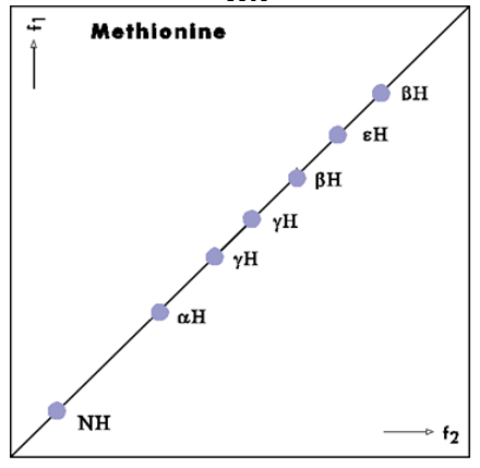
Question: Shown in the first figure is the diagonal for a COSY NMR spectrum for a methionine residue. (Assume the methionine is in the middle of
- Shown in the first figure is the diagonal for a COSY NMR spectrum for a methionine residue. (Assume the methionine is in the middle of a polypeptide chain, and the peaks here represent an isolated spin system.) Recall that the diagonal of the COSY spectrum corresponds to the one-dimensional proton NMR spectrum.
- Draw the chemical structure of methionine and label the N, , , , and protons on your drawing.
(Note that there are two protons and two protons that appear to be in different chemical environments and thus have different chemical shifts.)
- Draw the expected cross peaks on the COSY spectrum. (You only need to draw the cross peaks above or below the diagonal, as the spectrum is symmetric.) Explain your thinking
- Shown on the next figure is the diagonal for a NOESY spectrum for a methionine-alanine dipeptide. The methionine peaks are the same as in the previous question; the peaks due to the alanine protons are drawn in red.
- Draw the expected cross peaks in this spectrum, assuming the dipeptide is in an extended conformation. Explain your thinking
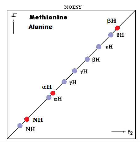
- Draw the expected cross peaks in this spectrum, assuming the dipeptide is in an extended conformation. Explain your thinking
f1 Methionine H H H H NH f2 NOESY ; Methionine Alanine H H H H H NH NH f2
Step by Step Solution
There are 3 Steps involved in it
1 Expert Approved Answer
Step: 1 Unlock

Question Has Been Solved by an Expert!
Get step-by-step solutions from verified subject matter experts
Step: 2 Unlock
Step: 3 Unlock