

Tales from the grave: Opposing autopsy reports from a body exhumed 1. Introduction Exhumations in criminal cases
Question:
Tales from the grave: Opposing autopsy reports from a body exhumed
1. IntroductionExhumations in criminal cases are rare, but on occurrence they can resolve issues that are either overlooked or unknown during an initial investigation.1 An unknown issue can manifest itself through facts gleaned by further investigation or by advances in the forensic sciences, of which DNA analysis is the latest example. Most exhumations are the result of court orders, when the specific cause of death remains an issue.1e3 With skeletonisation taking anywhere between 4 and 10 years, a high rate of forensic clarification of the cause of death can be gained from exhumations even after years of burial.1e3
Toxicology tests can often shed necessary light on a case in which suspicious death has occurred in drug-related cases.3 However, illegal drug toxicity, such as cocaine toxicity, can be relatively difficult to determine as the specific cause of death when combined with various types of physical trauma because it may be difficult to attribute the cause of death to one particular bodily injury.4 Thus questions arise. Did the illegal drug toxicity alone cause death? Did the physical trauma alone cause death? Did
* Corresponding author. Tel.: þ1 (281) 275 8826; fax: þ1 (281) 275 3361. E-mail address: GunasekeraR@uhv.edu (R.S. Gunasekera). |
a combination of illegal drug toxicity and physical trauma cause death? If so, what is the proportionate causation?
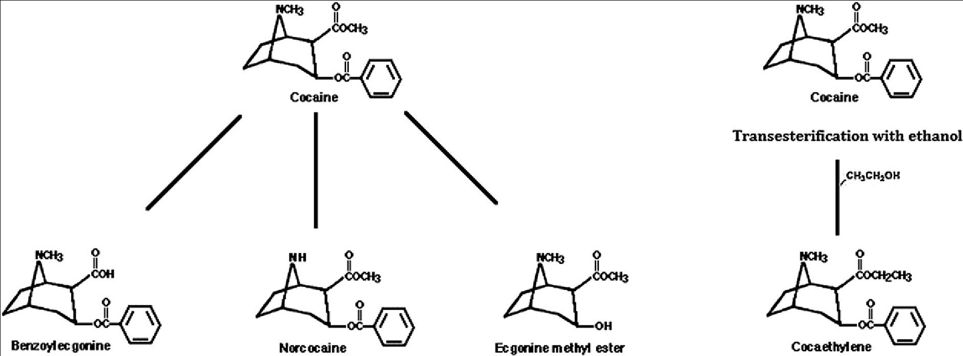
In addition to the analytical complexities created by intervening non-drug-relatedfactors,forensicallyconcludingthat adrugoverdose is the specific cause of a death itself is complicated by biological and toxicological factors. Cocaine provides an excellent example of the complexities that can arise. First, cocaine can be ingested by various ways: inhaled as a vapour, snorted or injected intravenously.5 The method of ingestion affects the rapidity with which the drug shocks and otherwise affects the body. Second, cocaine toxicity can affect the body in different ways to different degrees, depending on one’s physical condition and congenital tolerance. Cocaine is a nervous system stimulant that prevents the re-uptake of dopamine, serotonin andnorepinephrine intothepresynapticneuronsbindingtotransport proteins (Fig. 1).6 With excessive use, ischaemic heart disease can develop.5 Cardiotoxicity occurs with prolonged drug use and is the cause of most cocaine-related sudden deaths.7 Rhabdomyolysis, intravascular coagulation, renal failure and convulsions can also occur.5 Hyperthermia is another symptom of toxicity that can lead to arrhythmias of the heart and hypertension.8 Third, because cocaine is metabolised differently in each individual, it is often impossible to determine with precision the quantity of the cocaine ingested.8
This is a case of a victim whose first autopsy determined her death to be an accidental overdose of cocaine. After 2 years, during
1752-928X/$ e see front matter 2012 Elsevier Ltd and Faculty of Forensic and Legal Medicine.
298 R.S. Gunasekera et al. / Journal of Forensic and Legal Medicine 19 (2012) 297e301
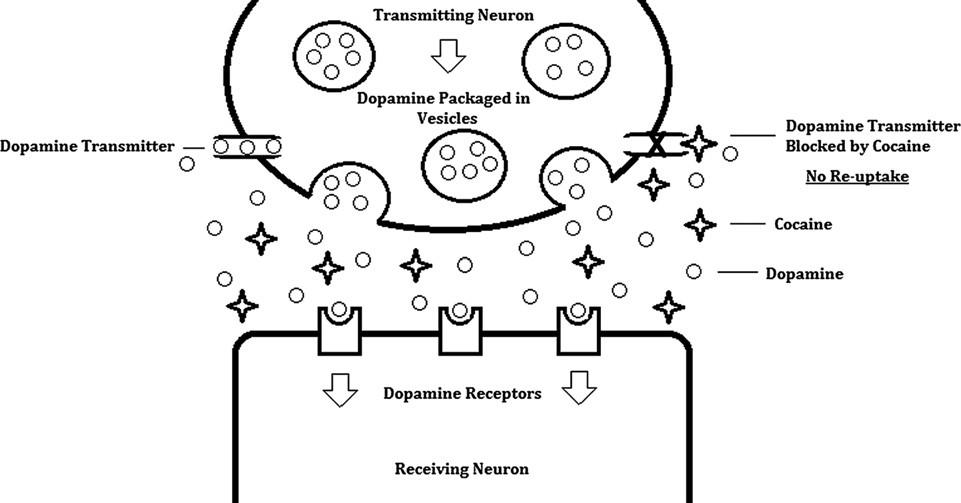
Fig. 1. The re-uptake of dopamine into the presynaptic neuron at the synapse.
which additional informationwas gathered bylaw enforcement, the victim was exhumed and a second autopsy was performed. The manner of death was changed to homicide with no specific cause.
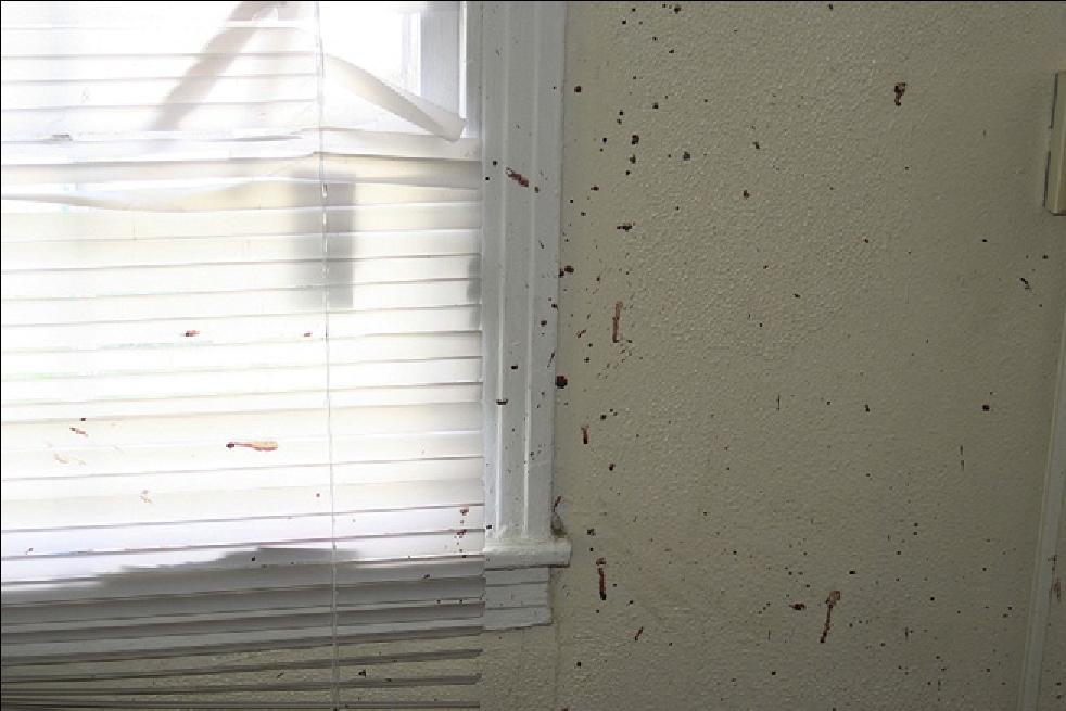
2. Case report2.1. Case descriptionIn March of 2006, a cleaning woman stumbled upon the body of a 42-year-old woman dead in her own apartment. The victim apparently had been deceased for multiple days and there was evidence of a struggle within her apartment. There was a trail of blood droplets in the threshold of her apartment leading down the common hallway of the complex, continuing up the stairs and ending in front of the apartment rented by suspect 2. Blood stains were found in the victim’s kitchen and in the kitchen sink as though someone had washed up in it. There was cast-off blood spatter, a bloodstain pattern created when blood is released or thrown from a blood-bearing object in motion, on the mini blinds above the sink and other blood spatter throughout the hallway leading to the bedroom and bathroom. In the bathroom, blood was found in the sink and tub and a pair of men’s blood-stained blue jeans was also found. There was blood spatter on the door frame, walls and closet doorway in the bedroom where the victim was found lying on the floor between the bed and the wall (Fig. 2). There was a hole in the wall made with apparent force above the victim’s head (Fig. 3).
Statements made by an inmate at the local county jail and by others questioned by law enforcement brought the suspects into focus 2 months after the crime. The latent fingerprint report that was obtained also confirmed the presence of two of the suspects, which caused investigators to re-examine the body by requesting the courts for an exhumation.
3. Materials and methods3.1. DNA evidenceDNA was extracted from the crime scene evidence using the Chelex method of extraction.9 This was quantified using the Quantiblot Human DNA Quantitation Kit according to the manufacturer guidelines (Applied Biosystem, Foster City, CA.). DNA quantification was conducted on an ABI 7000 thermal cylcer and amplification was conducted on an ABI 9700 real-time thermal cycler. The cycling parameters for the amplification were 95 C for 11 min; 28 cycles of 94 C for 1 min, 59 C for 1 min, 72 C for 1 min; and a final extension at 60 C for 45 min.10,11 STR analysis was performed on the DNA evidence extracted using the AmpFlSTR Profiler Plus PCR Amplification and Typing Kit and the AmpFlSTR Cofiler PCR Amplification and Typing Kit (Applied Biosystems). PCR products were run by capillary electrophoresis with fluorescent detection with the ABI 310 and ABI 3130 genetic analysers (Applied Biosystems).
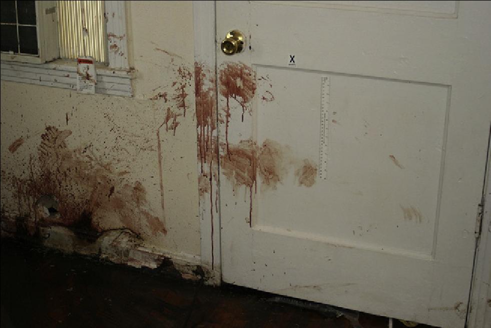
Fig. 2. Blood spatter in bedroom where body was found. Fig. 3. Hole in wall above victim’s head and blood evidence.
R.S. Gunasekera et al. / Journal of Forensic and Legal Medicine 19 (2012) 297e301 299
Table 1
Crime scene evidence submission and results.
Suspect 1 DNA evidence | |
Items matching Submission II, Item 10 (Stain A): Jacket, Item 32: Stain in kitchen, Item 42: Stain in hallway Submission V, Item 2 (Stain A): Jeans & belt. Submission VI, Item 31: Swab from instruction manual. | Submission V, Item 1 (Stain A)–blue pants in bathroom |
Probability of match Probability: 1 in 515.5 quintillion for Caucasian (C), 1 in 101.6 Blacks (B), and 1 in 8.299 sextillion for Hispanics (H). Victim DNA evidence | 1 in 399.8 quadrillion for C, 1 in 50.63 quadrillion for B, and 1 in 2.872 quintillion for H. |
Items matching Submission V, Item 1 (Stain B): Blue pants, Item 2B (Stain A): white long john pants. Mixture of Suspect 1 and victim DNA evidence | Submission V, Item 4: Swab from bathtub. |
Items matching Submission V, Item 5: Swab from sink. | Submission V, Item 6: Kitchen knife. |
Probability of match to 1 in 121.3 million for C, 1 in 14.61 million for B, and Suspect 1 1 in 768 million for H. | 1 in 40.63 billion for C, 1 in 2.86- billion for B, and 1 in 67.75 billion for H. |
DNA from suspect 1 was found on items of clothing and in the bathtub and sinks, and mixtures of the victim’s blood and suspect 1’s blood were found in the bathtub and sink, as well as on clothing and the kitchen knife thought to be used in the crime (Table 1).
4.2. Fingerprint evidenceLatent fingerprints found on a beer can, an appliance instruction manual and the kitchen knife also matched suspect 1. Fingerprints found on a white plastic bag matched suspect 2 placing him in the apartment on the night of the crime.
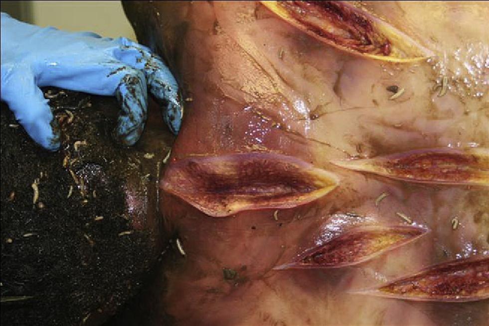
4.3. First autopsy findingsIt was noted at the initial autopsy that the preservation of the body was poor and there was advanced decomposition artefact with dark and tan colouration and extensive skin slip, the result of being enclosed in a room with no ventilation during intense summer heat. No lividity or rigidity of the extremities was observed. There were no injuries to the hands. The back and buttocks had dark colouration with bruising over the top of the scapula and one bruise over each lower scapula (Fig. 4). Lacerations were found on the bridge of the nose and left cheek. There was a defect over the bridge of the nose that extended to the forehead and followed the contour of the medial right eye. It extended downward to the right naso-orbital fold. A second defect was found
on the left face that followed the contour of the left zygomatic arch. The scalp, skull and dura showed no remarkable findings and the intracranial contents (675 g) were completely liquefied. No focal haemorrhage in the epidural, subdural and intraparenchymal space were found (Fig. 5). The trunk was opened and no haemorrhage or abnormal fluids were found. The bony, cartilaginous and soft tissues of the neck were without trauma. Examination of the heart found a normal size (274 g) and configuration with normal distribution of the coronary arteries with the right dominant, patent, and without significant atherosclerosis. No injury to the myocardium, the foramen ovale was closed and the septae were intact. All valves were thin, delicate and flexible and the aorta was intact with mild atherosclerosis of the intimal surface. The lungs were normal and unremarkable as were the liver and gall bladder. The stomach contained decomposed blood to a point approximately 4 inches into the duodenum. No drug remnants such as tablets or capsules were found within the gastric contents. The uterus is notable for a 2-cm-solid leiomyoma with a whorled white solid cut surface. There were no abnormalities of the axial and appendicular skeletal systems and the skeletal muscles were unremarkable. Toxicology results showed the presence of cocaine metabolites. In the muscle, gas chromatographemass spectrometry (GCeMS) showed that concentrations of cocaine were 0.77 mg l1, cocaethylene was detected and ethanol was 0.04%. In the bile, cocaine and cocaethylene were detected. Since there was no apparent lifethreatening injury, and due to the high (toxic) levels of cocaine and cocaine metabolites in tissues, a cause of death was determined to be cocaine overdose and the manner of death was determined to be an accident. The medical examiner (ME) in this initial autopsy was not provided with crime scene data. It is not customary for
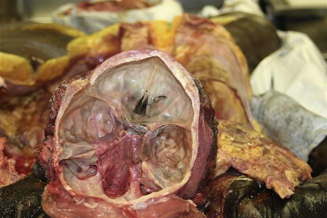
Fig. 4. Bruising and lacerations of scapula.
Fig. 5. Skull with no focal hemorrhage in the epidural, subdural, and intraparenchymal space.
300 R.S. Gunasekera et al. / Journal of Forensic and Legal Medicine 19 (2012) 297e301
a medical examiner to visit the crime scene and usually the ME’s office depends on the crime scene investigators (CSIs) to provide such information to the appropriate authorities. The ME thus did not have the information regarding the physical findings at the scene and only had information found within the body to make an opinion. This led to a finding of death due to cocaine overdose due to elevated levels of the drug and its metabolites found in the toxicology results.
4.4. Second autopsy findingsThe second autopsy was performed in February of 2008, approximately 2 years after the first autopsy, and confirmed much of the first autopsy along with additional findings. The body was in a Ziegler box secure with screws and enclosed in a blue body bag, which was within a white body bag, which was wrapped in a blue plastic sheet. The body was not embalmed.
The additional findings were injury related. Upon further examination of the lacerations of the face, it was found that the right side of the nasal bone was fractured off in a curvilinear manner (Fig. 6). There was an irregular jagged border of the medial aspect of the left side of the nasal bone including a small area that was likely fractured off. The same was found of the nasal bone near the bridge of the nose. Fractures were found in the inferior aspect of each orbit, greater in the left than the right (Fig. 7). On the right side near the back of the orbit, there was a 0.4-cm piece of bone in the inferior aspect of the orbit that was fractured and displaced posteriorly/inferiorly. It was aligned with a fracture in the more anterior orbit. The skin, subcutaneous tissue and skeletal muscle of both arms from the hands to the shoulders were reflected along with those of the back, posterior thighs and buttocks. The arms showed no discolouration or convincing blood extravasation except for the left wrist and distal forearm, and the lateral aspect of the left shoulder. These had large areas of discolouration that could have been blood extravasation or contusion. The back, legs and buttocks do not show any convincing areas of discolouration or blood extravasation. The second autopsy stated that death was due to homicide given the totality of evidence.
5. DiscussionThe conclusion of the first autopsy was made cautiously because of the lack of information regarding the circumstances surrounding the death. There was no obvious evidence of life-threatening trauma; body decomposition had been rapid. Therefore, the only reasonable probable medical cause of death at the time of the first autopsy was the elevated levels of cocaine and cocaethylene shown
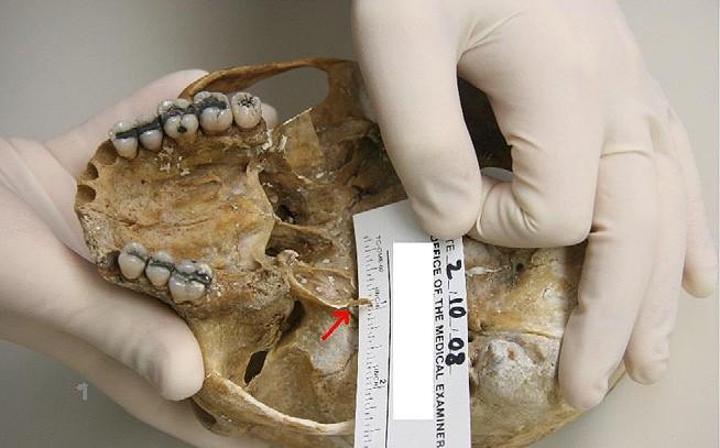
Fig. 6. Curvilinear fracture of the nasal bone. (Inferior view of the basal skull depicting orbital and nasal bones).
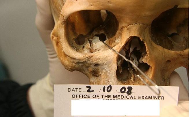
Fig. 7. Fractures found in the inferior aspect of each orbit and nasal bone.
in the toxicology report. Chronic cocaine users often exhibit decreased body mass index (BMI) and increased heart weight because of the effect cocaine has on the metabolism of serotonergic neurons, which are involved in appetite control.12 The victim in this case did not exhibit either of these signs of chronic use. Furthermore, there was no evidence of former cocaine-related stroke in the brain, no evidence of heart issues related to the prolonged use of cocaine, and no evidence of other physical traumas related to cocaine use.12 The organs presented normally. These findings substantiate previous reports of cocaine toxicity being difficult to determine definitively without further physical evidence and reports from witnesses and investigators.4,5,7,8
Cocaine is rapidly converted to its hydrolytic products during life and undergoes quick bioconversion after death.6 Benzoylecgonine (BE) and ecgonine methyl ester (EME) are the most significant hydrolytic products of cocaine metabolism, and the biologically active ethyl trans-esterification analogue cocaethylene (CE) can be found in large amounts within the tissues of persons who also consume ethanol (Fig. 8).6 CE formation does not occur postmortem which suggests the active involvement of enzymes.6 The findings of EME and CE is an indication of oral use due to first-pass metabolism.6 In this case, cocaine, CE and ethanol were detected in the first toxicology report. This indicates that the victim did not die immediately after consuming the cocaine.
There are no doctrinal calculations or predetermined figures for what may constitute a lethal dose of a particular drug in postmortem toxicology because of the generally inherent lack of knowledge by the forensic examiner of certain information, such as the purity of the drug, how much drug was in the body at the instant of death and the number of doses taken over a particular period.4,8 Blood concentrations of the drug are different in death than in life and tolerance is different in each individual.4,8 Mode of ingestion is also important in determining toxicity. According to witness statements, the victim in this case ingested the cocaine through inhalation or smoking. Indeed, the number of variables highlights the importance of placing toxicological evidence into context with the general circumstances of the case, the autopsy findings and the findings of the criminal investigator.
R.S. Gunasekera et al. / Journal of Forensic and Legal Medicine 19 (2012) 297e301 301
Fig. 8. Metabolism of cocaine. |
Exhumations can be extraordinarily useful in criminal cases where contradicting evidence comes to light and additional physical evidence is needed to find the truth. Subsequent autopsies have been helpful in revealing the cause of death many years after burial. In this case, some 2 years after the victim had been buried, police investigators had continued to gather additional information surrounding the circumstances of death. Later during a second medical examination, the forensic pathologist uncovered wounds indicating a homicide. Possessed of information giving context to the victim’s death, the pathologist found fractures to the victim’s face from the beating she received, in the second autopsy. These findings added physical evidence to the hypothesis of homicide. Interestingly, the second medical examiner determined some of the facial wounds to be the result of a sharp instrument, but neither medical examiner reported finding defensive wounds to the hands or arms. However, defensive injuries are not always seen in an attack with a sharp instrument.13 In their study of sharp force injuries, Schmidt and Pollak observed defence injuries in only 45.9% of their subjects.13 In this case, physical evidence was put into context with witness testimony and other investigative evidence, such as STR analysis and fingerprint analysis.
Suspect 1 was linked to the crime through DNA evidence, latent fingerprintsandwitnesstestimony. Hereceivedlifeinprison.Suspect 2 was linked to the crime by latent fingerprint evidence, witness testimony and a confession. He received 45 years in prison. Suspect 3 was linked only by testimony. She received 20 years in prison.
This case is significant because it demonstrates how forensic analysis conducted in relation to the varying quality of information surrounding the circumstances of a violent death can lead two medical examiners acting independently of each other to reach different conclusions. The case also demonstrates the difficulties that can arise when attempting to determine the cause of death in criminal circumstances, for death can result from one or more possible independent causes, or a combination thereof. The subject case highlights that the level of cocaine toxicity is not easily determined in putrefied tissue because of numerous physiological variables, including genetic predisposition, BMI, metabolism and biochemistry. Although exhumations can have a place in providing forensic information in a criminal investigation, this case suggests that bodies that have undergone advanced decomposition leading to skeletonisation may provide only limited evidence in determining cause of death. Indeed, in advanced decomposition, injuries are known to be lost and/or be altered, making it difficult to deduce any conclusion for the cause of death.14 It is noteworthy to mention that the outcome of such cases should be carefully concluded with comparative reference and careful study of similar cases in criminal history. Nonetheless, despite leaving some medical issues unresolved, the contradictory conclusions of medical examiners are useful in examining the circumstances leading to death in criminal circumstances.
Expert Answer:
Introduction Exhumations in criminal cases are rare but on occurrence they can resolve issues that are either overlooked or unknown during an initial investigationAn unknown issue can manifest itself ... View the full answer


Smith and Roberson Business Law
ISBN: 978-0538473637
15th Edition
Authors: Richard A. Mann, Barry S. Roberts
