

Question: The tissue slice being imaged by a parallel beam x-ray CT scanner is f(x,y)=rect(x/3,y+1/2)+rect(x,y). (a) Assume the detector is a point detector. Sketch the projection
The tissue slice being imaged by a parallel beam x-ray CT scanner is f(x,y)=rect(x/3,y+1/2)+rect(x,y). (a) Assume the detector is a point detector. Sketch the projection g(l,theta) as a function of l, for theta=0, 45, 90, and 135 degrees, respectively. You should indicate the magnitudes of the projected values where necessary on your sketch. (b) Sketch the image obtained by backprojections from both 0 and 90 degree projections. You should normalize your back-projection using the dimension of the imaged region as indicated on the figure. (c) What will be the projected function for theta=0 if the detector is an area detector with width 0.1 cm. Sketch the projected function. (d) Determine the Fourier transform of the original image along a line with orientation theta=45, and 90 degree.
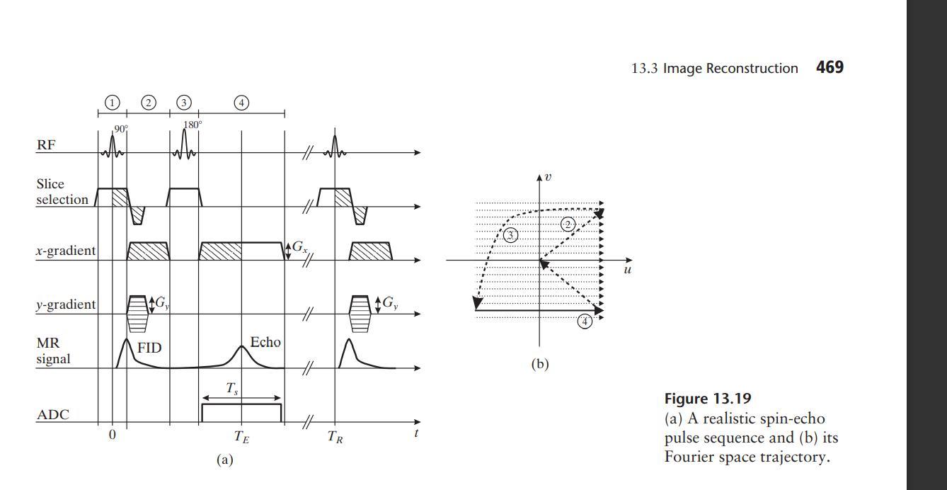
RF Slice selection x-gradient y-gradient MR signal ADC ,90% G FID 180 T (a) Echo TE TR AG, AV (b) 13.3 Image Reconstruction 469 u Figure 13.19 (a) A realistic spin-echo pulse sequence and (b) its space trajectory. Fourier
Step by Step Solution
3.38 Rating (167 Votes )
There are 3 Steps involved in it
Iran I rom Ay ... View full answer

Get step-by-step solutions from verified subject matter experts