

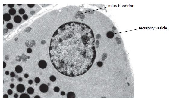
The electron micrograph on page 25 shows part of a secretory cell from the pancreas. The secretory
Question:
The electron micrograph on page 25 shows part of a secretory cell from the pancreas. The secretory vesicles are Golgi vesicles and appear as dark round structures. The magnification is × 8000.
a. Copy and complete the table. Use a ruler to help you find the actual sizes of the structures. Give your answers in micrometres.
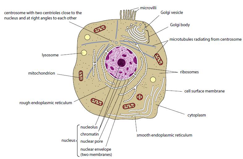
b. Make a fully labelled drawing of representative parts of the cell. You do not have to draw everything, but enough to show the structures of the main organelles. Use a full page of plain paper and a sharp pencil. Use Figures 1.16 and 1.17 in this book and the simplified diagram in d below to help you identify the structures.
c. The mitochondria in pancreatic cells are mostly sausage-shaped in three dimensions. Explain why some of the mitochondria in the electron micrograph below appear roughly circular.
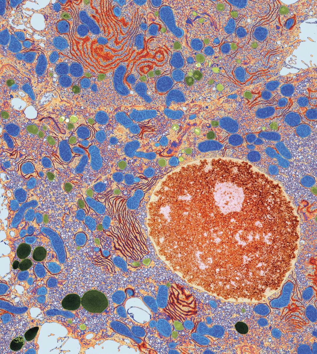
The figure below shows a diagram based on an electron micrograph of a secretory cell from the pancreas. This type of cell is specialised for secreting (exporting) proteins. Some of the proteins are digestive enzymes of the pancreatic juice. The cell is very active, requiring a lot of energy. The arrows show the route taken by the protein molecules.

i. Describe briefly what is happening at each of the stages A, B, C and D.
ii. Name one molecule or structure which leaves the nucleus by route E.
iii. Through which structure must the molecule or structure you named in ii pass to get through the nuclear envelope?
iv. Name the molecule which leaves the mitochondrion in order to provide energy for this cell.
Figures 1.16
Figures 1.17
Step by Step Answer:
Typical animal cell 10 20 angstrom in diameter which is about onefifth the size of the smallest particle p is equal to the next it was not until good ...View the full answer


Cambridge International AS And A Level Biology
ISBN: 9781107636828
4th Edition
Authors: Mary Jones, Richard Fosbery, Jennifer Gregory, Dennis Taylor
