Because of intravascular resistance to oxygen diffusion, the oxygen partial pressure at the blood-tissue interface may be
Question:
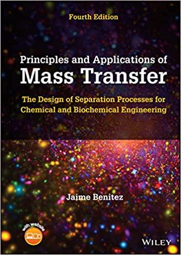
Because of intravascular resistance to oxygen diffusion, the oxygen partial pressure at the blood-tissue interface may be less than the average oxygen partial pressure in the blood. This effect can be represented approximately by the expression (McGuire and Secomb, 2001):
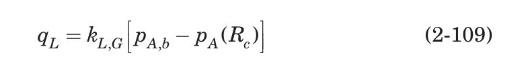
where \(q_{L}\) is the rate of oxygen diffusion from the capillary into the surrounding tissue per unit length of capillary, and \(k_{L, G}\) is an intravascular mass-transfer coefficient. For this case, a Sherwood number has been defined as
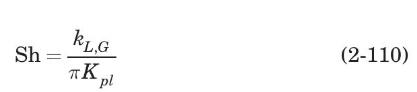
where \(K_{p l}\) is the Krogh oxygen diffusion constant in plasma \(=8.3 \times 10^{-10} \mathrm{~cm}^{3}\) of \(\mathrm{O}_{2} / \mathrm{cm} \cdot \mathrm{s} \cdot\) torr. The value of Sh depends on the vessel diameter. For example, for a capillary with a 5 - \(\mu \mathrm{m}\) diameter, \(\mathrm{Sh}=2.5\) (McGuire and Secomb, 2001).
(a) Show that, for the Krogh cylinder model described in Example 1.28,
(b) For the conditions described in Example 1.28, at the capillary entrance, calculate the oxygen partial pressure at the capillary wall.
Data From Example 1.28:-
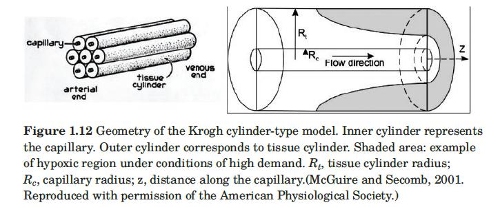
In 1922, August Krogh proposed that oxygen was transported from capillaries to surrounding tissue by passive diffusion. He formulated a simple geometric model to describe this phenomenon. The Krogh model is a simplification of a capillary bed; a group of parallel identical units, each containing a central capillary and a surrounding cylinder of tissue, as shown in Figure 1.12. The goal of the Krogh model is to predict the concentration of oxygen as a function of position within the tissue cylinder. Each tissue cylinder is assumed to be supplied with oxygen exclusively by the capillary within it. Oxygen diffusion in the axial direction is neglected, as evidenced by the fact that the oxygen concentration gradient is much steeper in the radial direction than in the axial direction. Applying Fick’s law of diffusion in the radial direction (Rc ≤ r ≤ Rt) and conservation of mass lead to the following equation for oxygen diffusion in muscle tissue (McGuire and Secomb, 2001):
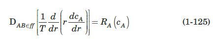
where ca is the oxygen concentration in tissue. In studies of oxygen transport, it is customary to express the concentration in tissue in terms of an equivalent oxygen partial pressure, pa = HcA, where H is a modified form of Henry’s law constant. Then, equation (1-125) can be written in terms of the oxygen partial pressure in tissue as

where K (known as the Krogh diffusion coefficient) = DAB,eff/H, and M(pA) is the oxygen consumption rate per unit volume of the tissue cylinder. Oxygen consumption in skeletal muscle is usually assumed to follow Michaelis-Menten kinetics, a model that describes the kinetics of many enzymatic reactions, given by
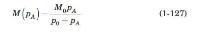
where M0 is the oxygen demand (i.e., the consumption when the oxygen supply is not limiting) and p0 is the partial pressure of oxygen when consumption is half of the demand. The Michaelis-Menten equation reduces to first-order kinetics for values of pA much smaller than p0, and to zero-order kinetics for values of pA much larger than p0. For this example, we will assume zero-order kinetics; therefore, M(pA) = M0.
The two boundary conditions needed to solve equation (1-126) are i) Neglecting the oxygen-diffusion resistance of the capillary wall, at the capillary–tissue interface, the oxygen partial pressure in the tissue is approximately equal to the average partial pressure of oxygen within the blood, PA b; that is, PA(RC) = PA,b.
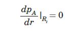
ii) It is assumed that no oxygen is exchanged across the outer boundary of the tissue cylinder, so that
The complete solution for zero-order kinetics is
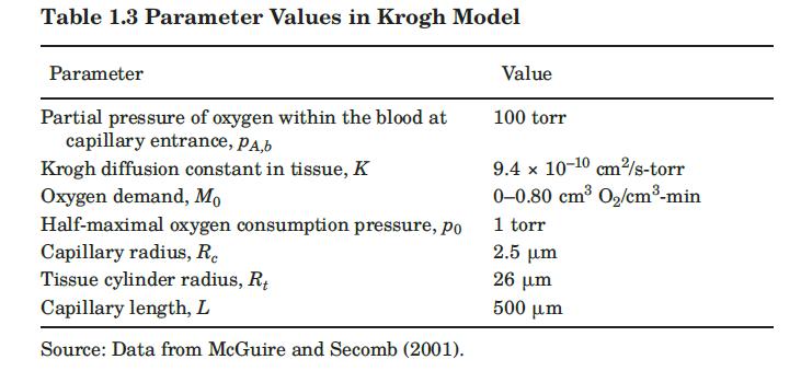
Equation (1-128) can be used to predict whether, under a given set of parameters, there are hypoxic regions—where the partial pressure of oxygen is less than 1 torr— in the tissue cylinder. Table 1.3 summarizes parameter values used in the Krogh model.
To illustrate the use of equation (1-128), we use the parameters of Table 1.3, with an oxygen consumption of 0.4 cm3 O2/cm3-min, and estimate that the lowest oxygen partial pressure in the tissue surrounding the capillary entrance is 49.9 torr at r = Rt. Therefore, all the tissue at this position is exposed to a healthy oxygen concentration with no hypoxic regions. Moreover, since pA is everywhere much higher than p0, the assumption of zero-order kinetics is validated.
Step by Step Answer: