

Question: Using Section 3.3 as a reference, suppose you want to observe the surface of a microscopic section of bone. Would the best choice for this
Using Section 3.3 as a reference, suppose you want to observe the surface of a microscopic section of bone. Would the best choice for this task be a compound light microscope or an electron microscope?
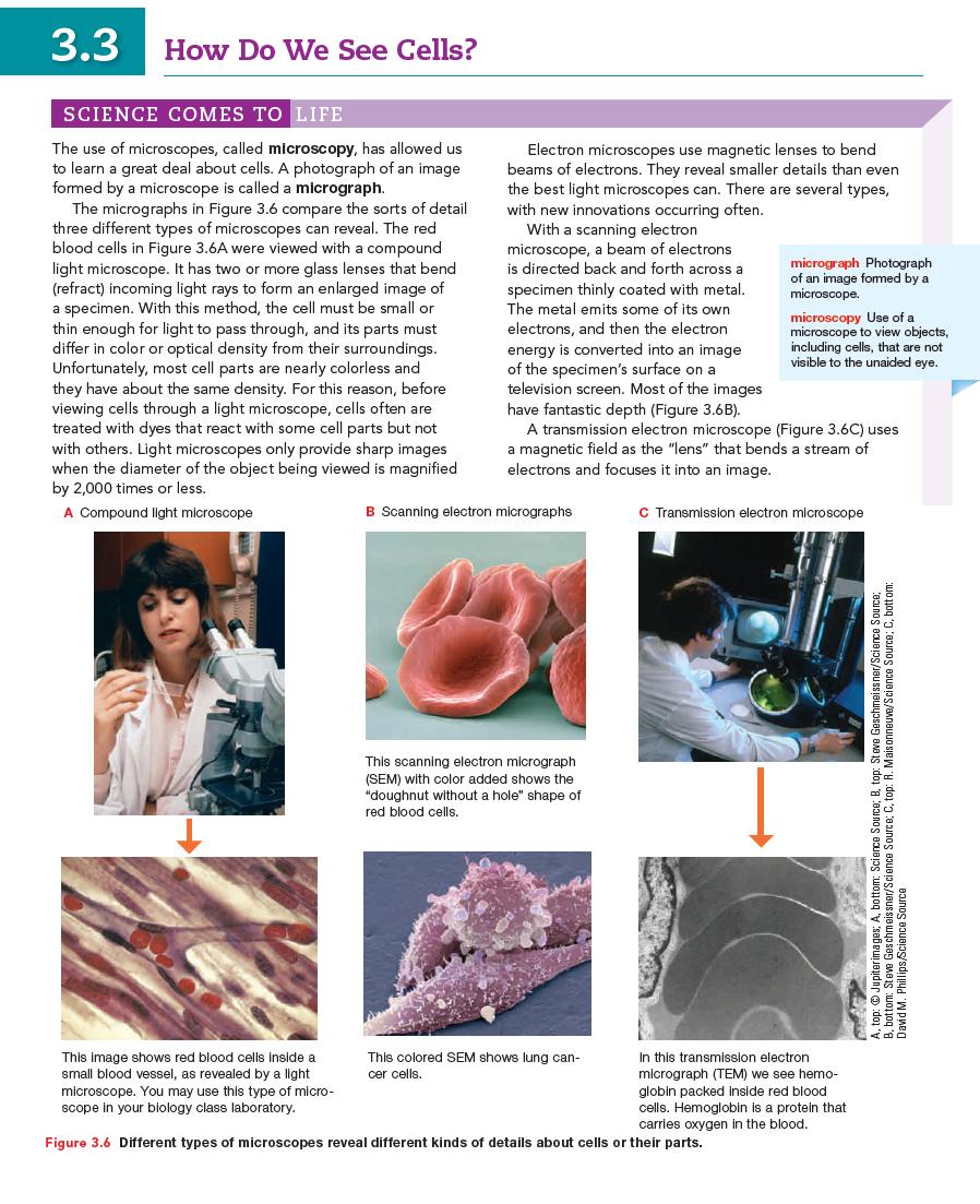
3.3 How Do We See Cells? SCIENCE COMES TO LIFE The use of microscopes, called microscopy, has allowed us to learn a great deal about cells. A photograph of an image formed by a microscope is called a micrograph. The micrographs in Figure 3.6 compare the sorts of detail three different types of microscopes can reveal. The red blood cells in Figure 3.6A were viewed with a compound light microscope. It has two or more glass lenses that bend (refract) incoming light rays to form an enlarged image of a specimen. With this method, the cell must be small or thin enough for light to pass through, and its parts must differ in color or optical density from their surroundings. Unfortunately, most cell parts are nearly colorless and they have about the same density. For this reason, before viewing cells through a light microscope, cells often are treated with dyes that react with some cell parts but not with others. Light microscopes only provide sharp images when the diameter of the object being viewed is magnified by 2,000 times or less. A Compound light microscope This image shows red blood cells inside a small blood vessel, as revealed by a light microscope. You may use this type of micro- scope in your biology class laboratory. Electron microscopes use magnetic lenses to bend beams of electrons. They reveal smaller details than even the best light microscopes can. There are several types, with new innovations occurring often. With a scanning electron microscope, a beam of electrons is directed back and forth across a specimen thinly coated with metal. The metal emits some of its own electrons, and then the electron energy is converted into an image of the specimen's surface on a television screen. Most of the images have fantastic depth (Figure 3.6B). A transmission electron microscope (Figure 3.6C) uses a magnetic field as the "lens" that bends a stream of electrons and focuses it into an image. B Scanning electron micrographs This scanning electron micrograph (SEM) with color added shows the "doughnut without a hole" shape of red blood cells. This colored SEM shows lung can- cer cells. micrograph Photograph of an image formed by a microscope. microscopy Use of a microscope to view objects, including cells, that are not visible to the unaided eye. C Transmission electron microscope Figure 3.6 Different types of microscopes reveal different kinds of details about cells or their parts. In this transmission electron micrograph (TEM) we see hemo- globin packed inside red blood cells. Hemoglobin is a protein that carries oxygen in the blood. Source; top:Jupiterimages. A bottom: Science Source; B, top: Steve Geschmeissner/Science David M. Phillips/Science Source Source; C. top: H. Mais onneuve/Science Source; C, bottom: SPEEES
Step by Step Solution
3.36 Rating (149 Votes )
There are 3 Steps involved in it
To observe the surface of a microscopic section of bone the best choice would be a scanning electron microscope SEM rather than a compound light micro... View full answer

Get step-by-step solutions from verified subject matter experts