

Question: Can you found coding from qusetion for BICD-10? 13 The patient is contrast. The clinical muscle weaknes and in the left shift of the midis
Can you found coding from qusetion for BICD-10? 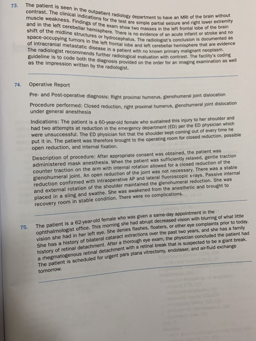
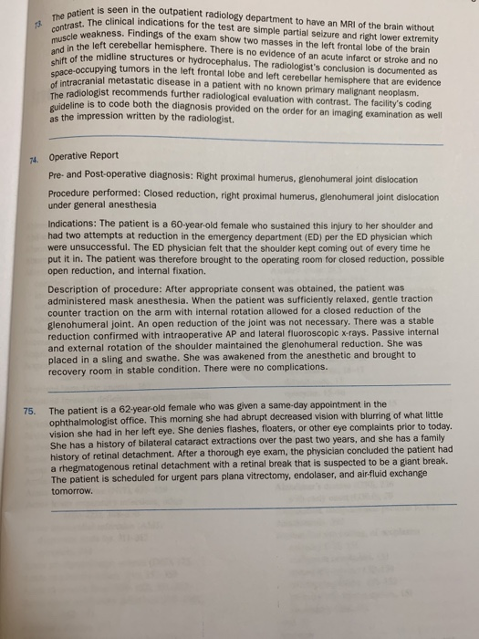
13 The patient is contrast. The clinical muscle weaknes and in the left shift of the midis + is seen in the outpatient radiology department to have an MRI of the brain without The clinical indications for the test are simple partial seizure and right lower extremity weakness. Findings of the exam show two masses in the left frontal lobe of the brain the left cerebellar hemisphere. There is no evidence of an acute infarct or stroke and no the midline structures or hydrocephalus. The radiologist's conclusion is documented as Secupying tumors in the left frontal lobe and left cerebellar hemisphere that are evidence acranial metastatic disease in a patient with no known primary malignant neoplasm. radiologist recommends further radiological evaluation with contrast. The facility's coding Weline is to code both the diagnosis provided on the order for an imaging examination as well as the impression written by the radiologist. space-occupying tumor 14. Operative Report Pre- and Post-operative diagnosis: Right proximal humerus, glenohumeral Joint dislocation Procedure performed: Closed reduction, right proximal humerus, glenohumeral joint dislocation under general anesthesia Indications: The patient is a 60-year-old female who sustained this injury to her shoulder and had two attempts at reduction in the emergency department (ED) per the ED physician which were unsuccessful. The ED physician felt that the shoulder kept coming out of every time he put it in. The patient was therefore brought to the operating room for closed reduction, possible open reduction, and internal fixation. Description of procedure: After appropriate consent was obtained, the patient was administered mask anesthesia. When the patient was sufficiently relaxed, gentle traction counter traction on the arm with internal rotation allowed for a closed reduction of the glenohumeral joint. An open reduction of the joint was not necessary. There was a stable reduction confirmed with intraoperative AP and lateral fluoroscopie x-rays. Passive internal and external rotation of the shoulder maintained the glenohumeral reduction. She was placed in a sling and swathe. She was awakened from the anesthetic and brought to recovery room in stable condition. There were no complications. The patient is a 62-year-old female who was given a same-day appointment in the ophthalmologist office. This morning she had abrupt decreased vision with blurring of what little vision she had in her left eye. She denies flashes, floaters, or other eye complaints prior to today. She has a history of bilateral cataract extractions over the past two years, and she has a family history of retinal detachment. After a thorough eye exam, the physician concluded the patient had a rhegmatogenous retinal detachment with a retinal break that is suspected to be a giant break. The patient is scheduled for urgent pars plana vitrectomy, endolaser, and air-fluid exchange tomorrow. The patient is seen in the outpatient radiology department to have a contrast. The clinical indications for the test are simple partial sex muscle weakness. Findings of the exam show two masses in and in the left cerebellar hemisphere. There is no evidence of an a shift of the midline structures or hydrocephalus. The radiolo space-occupying tumors in the left frontal lobe and left cerebellar h of intracranial metastatic disease in an The radiologist recommends further radiological evaluation with contrast artment to have an MRI of the brain without Simple partial seizure and right lower extremity am show two masses in the left frontal lobe of the brain no evidence of an acute infarct or stroke and no mydrocephalus. The radiologist's conclusion is documented as disease in a patient with no known primary malignant neoplasm left cerebellar hemisphere that are evidence guideline is to code both the sto code both the diagnosis provided on the order for an imaging examinatio evaluation with contrast. The facility's coding in the order for an imaging examination as well as the impression written by the radiologist. 74. Operative Report Pre- and Post-operative diagnosis: Right proximal humerus, glenohumeral joint dislocation Procedure performed: Closed reduction, right proximal humerus, glenohum under general anesthesia Indications: The patient is a 60-year-old female who sustained this injury to her shoulder and had two attempts at reduction in the emergency department (ED) per the ED physician which were unsuccessful. The ED physician felt that the shoulder kept coming out of every time he put it in. The patient was therefore brought to the operating room for closed reduction, possible open reduction, and internal fixation. Description of procedure: After appropriate consent was obtained, the patient was administered mask anesthesia. When the patient was sufficiently relaxed, gentle traction counter traction on the arm with internal rotation allowed for a closed reduction of the glenohumeral joint. An open reduction of the joint was not necessary. There was a stable reduction confirmed with intraoperative AP and lateral fluoroscopic x-rays. Passive internal and external rotation of the shoulder maintained the glenohumeral reduction. She was placed in a sling and swathe. She was awakened from the anesthetic and brought to recovery room in stable condition. There were no complications. 75. She has a history of bilatert After a thorough eye exam, the prys The patient is a 62-year-old female who was given a same day appointment in the ophthalmologist office. This morning she had abrupt decreased vision with blurring of what little vision she had in her left eye. She denies tlashes, Floaters, or other eye complaints prior to today. She has a history of bilateral cataract extractions over the past two years, and she has a family history of retinal detachment. After a thorough eye exam, the physician concluded the patient had arhegmatogenous retinal detachment with a retinal break that is suspected to be a giant break The patient is scheduled for urgent pars plana vitrectomy, endolaser, and air-fluid exchange tomorrow. 13 The patient is contrast. The clinical muscle weaknes and in the left shift of the midis + is seen in the outpatient radiology department to have an MRI of the brain without The clinical indications for the test are simple partial seizure and right lower extremity weakness. Findings of the exam show two masses in the left frontal lobe of the brain the left cerebellar hemisphere. There is no evidence of an acute infarct or stroke and no the midline structures or hydrocephalus. The radiologist's conclusion is documented as Secupying tumors in the left frontal lobe and left cerebellar hemisphere that are evidence acranial metastatic disease in a patient with no known primary malignant neoplasm. radiologist recommends further radiological evaluation with contrast. The facility's coding Weline is to code both the diagnosis provided on the order for an imaging examination as well as the impression written by the radiologist. space-occupying tumor 14. Operative Report Pre- and Post-operative diagnosis: Right proximal humerus, glenohumeral Joint dislocation Procedure performed: Closed reduction, right proximal humerus, glenohumeral joint dislocation under general anesthesia Indications: The patient is a 60-year-old female who sustained this injury to her shoulder and had two attempts at reduction in the emergency department (ED) per the ED physician which were unsuccessful. The ED physician felt that the shoulder kept coming out of every time he put it in. The patient was therefore brought to the operating room for closed reduction, possible open reduction, and internal fixation. Description of procedure: After appropriate consent was obtained, the patient was administered mask anesthesia. When the patient was sufficiently relaxed, gentle traction counter traction on the arm with internal rotation allowed for a closed reduction of the glenohumeral joint. An open reduction of the joint was not necessary. There was a stable reduction confirmed with intraoperative AP and lateral fluoroscopie x-rays. Passive internal and external rotation of the shoulder maintained the glenohumeral reduction. She was placed in a sling and swathe. She was awakened from the anesthetic and brought to recovery room in stable condition. There were no complications. The patient is a 62-year-old female who was given a same-day appointment in the ophthalmologist office. This morning she had abrupt decreased vision with blurring of what little vision she had in her left eye. She denies flashes, floaters, or other eye complaints prior to today. She has a history of bilateral cataract extractions over the past two years, and she has a family history of retinal detachment. After a thorough eye exam, the physician concluded the patient had a rhegmatogenous retinal detachment with a retinal break that is suspected to be a giant break. The patient is scheduled for urgent pars plana vitrectomy, endolaser, and air-fluid exchange tomorrow. The patient is seen in the outpatient radiology department to have a contrast. The clinical indications for the test are simple partial sex muscle weakness. Findings of the exam show two masses in and in the left cerebellar hemisphere. There is no evidence of an a shift of the midline structures or hydrocephalus. The radiolo space-occupying tumors in the left frontal lobe and left cerebellar h of intracranial metastatic disease in an The radiologist recommends further radiological evaluation with contrast artment to have an MRI of the brain without Simple partial seizure and right lower extremity am show two masses in the left frontal lobe of the brain no evidence of an acute infarct or stroke and no mydrocephalus. The radiologist's conclusion is documented as disease in a patient with no known primary malignant neoplasm left cerebellar hemisphere that are evidence guideline is to code both the sto code both the diagnosis provided on the order for an imaging examinatio evaluation with contrast. The facility's coding in the order for an imaging examination as well as the impression written by the radiologist. 74. Operative Report Pre- and Post-operative diagnosis: Right proximal humerus, glenohumeral joint dislocation Procedure performed: Closed reduction, right proximal humerus, glenohum under general anesthesia Indications: The patient is a 60-year-old female who sustained this injury to her shoulder and had two attempts at reduction in the emergency department (ED) per the ED physician which were unsuccessful. The ED physician felt that the shoulder kept coming out of every time he put it in. The patient was therefore brought to the operating room for closed reduction, possible open reduction, and internal fixation. Description of procedure: After appropriate consent was obtained, the patient was administered mask anesthesia. When the patient was sufficiently relaxed, gentle traction counter traction on the arm with internal rotation allowed for a closed reduction of the glenohumeral joint. An open reduction of the joint was not necessary. There was a stable reduction confirmed with intraoperative AP and lateral fluoroscopic x-rays. Passive internal and external rotation of the shoulder maintained the glenohumeral reduction. She was placed in a sling and swathe. She was awakened from the anesthetic and brought to recovery room in stable condition. There were no complications. 75. She has a history of bilatert After a thorough eye exam, the prys The patient is a 62-year-old female who was given a same day appointment in the ophthalmologist office. This morning she had abrupt decreased vision with blurring of what little vision she had in her left eye. She denies tlashes, Floaters, or other eye complaints prior to today. She has a history of bilateral cataract extractions over the past two years, and she has a family history of retinal detachment. After a thorough eye exam, the physician concluded the patient had arhegmatogenous retinal detachment with a retinal break that is suspected to be a giant break The patient is scheduled for urgent pars plana vitrectomy, endolaser, and air-fluid exchange tomorrow
Step by Step Solution
There are 3 Steps involved in it
1 Expert Approved Answer
Step: 1 Unlock

Question Has Been Solved by an Expert!
Get step-by-step solutions from verified subject matter experts
Step: 2 Unlock
Step: 3 Unlock