Question: Dance video 3-second segment dance sequence https://www.youtube.com/watch?v=TcWPiHjIExA Time stamp 1:03 to 1:06 Using knowledge from Chapters 1 and 2, discuss (12 sent.) the movements observed
Dance video 3-second segment dance sequence
https://www.youtube.com/watch?v=TcWPiHjIExA
Time stamp 1:03 to 1:06
Using knowledge from Chapters 1 and 2, discuss (12 sent.) the movements observed and the associated Joints (see table 1.2 and 2.3). (20pts) (quote the text when possible)
Select 10 DIFFERENT joints used in dance moves associated in the 3-second segment
Name of Joint
Type of Joint
Starting Position
Observed Joint Action
Plane of Motion
Axis of Motion
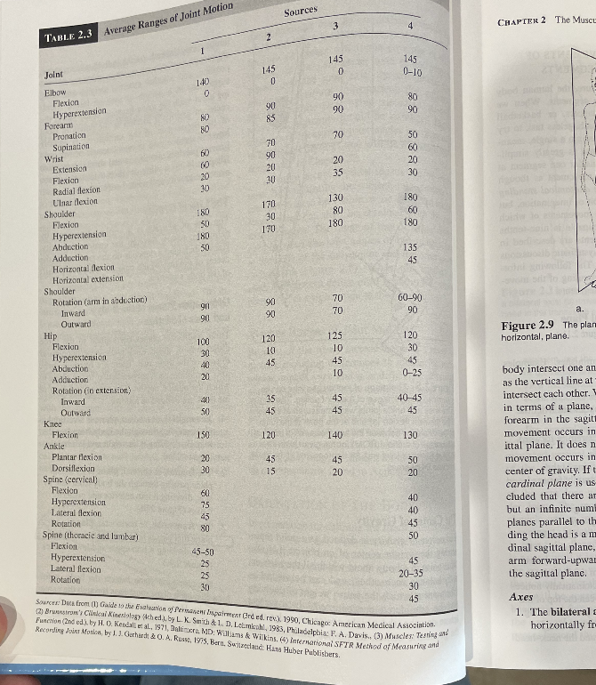
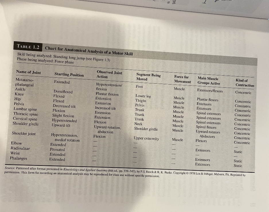
Sources Average Ranges of Joint Motion 3 4 CHAPTER 2 The Musc TABLE 2.3 1 145 145 Joint 145 0-10 140 Elbow Flexion 90 80 Hyperextension 90 90 Forearmi Promation 70 50 Supination 70 60 60 Wrist 90 20 20 Extension 20 35 30 Flexion Radial flexion LInar flexion 170 130 180 Shoulder 180 30 80 60 Flexion 170 180 180 Hyperextension 180 Abduction 50 135 Addiction 45 Horizontal flexion ofat anivolker Horizontal extension Shoulder Rotation (arm in abduction) 70 60-90 901 Inward 70 90 a 90 Outward Figure 2.9 The plan Hip 100 120 125 120 horizontal, plane. Flexion 10 30 Hyperextension 30 10 45 45 45 Abduction 40 10 body intersect one an Adduction 0-25 Rotation (in extension) as the vertical line at Inward 35 45 40-45 intersect each other.' Outward 50 45 45 in terms of a plane, Knee forearm in the sagit Flexion 150 120 140 130 movement occurs in Ankle ittal plane. It does n Plantar flexion 20 45 45 Dorsiflexion 50 movement occurs in 30 15 Spine (cervical) 20 20 center of gravity. If Flexion cardinal plane is us 60 Hyperextension 75 40 cluded that there an Lateral flexion 45 40 but an infinite num Rotation 45 planes parallel to th Spine [thoracic and lumbar ) 50 ding the head is a r Flexion 15-50 dinal sagittal plane Hyperextension 45 Lateral flexion 25 arm forward-upwa Rotation 25 20-35 the sagittal plane 30 45 Axes Sources: Data from (I) Gaide to the Evaluation y) Permanent Inpalmeat Ended. rev.). 1990, Chicago American Medical Association. 1. The bilateral 7Brunnstraw's Ciledeal Kinesiology (4ched.J. by L. K. Smith & 1. D. Lebamkohl, 1983, Philadelphia: F. A. Davis., (3) Muscles: Testing 813 Function (2nd ed.) by H. C. Kendall et al., 1971, Baltimore. MD: Williams & Wilkins. ( International SFTR Method of Measuring and horizontally fr Recording Jgiur Morlan, by J. J. Gerhard & O. A. Russe, 1975, Bern. Switzerland Hans Huber Publishers.PART I Anatomical and Physiological Fundamentals of Human Moll 24 TABLE 2.2 Approximate Ages of Epiphyseal Closures+ CHAPTER 2 The Musculosk TABLE 2.1 Approximate Ages of Epiphyseal Closures* Approximate Age Age while there is an increase 7-8 bone-eroding osteoclasts. SPINAL COLUMN Inferior rami of pubis and ischium almost change has been linked to h 25 complete occur with age, primarily in Vertebrae and sacrum 15-17 may include vitamin deficit THORAX 25 Upper extremity: scapula, lateral epicondyle of poor nutrition. In serious cas Sternum 25 weight bearing and muscle Ribs humerus, olecranon process of ulna UPPER EXTREMITY 18-19 some situations, falling may 25 Clavicle 15-17 Upper extremity: medial epicondyle of humans, breaking from their brittlen head and shaft of radius rosis negatively affects a pe Scapula Current research indicates Humerus 20 Lower extremity: femoral head and greater and Head fused with shaft 16-17 lesser trochanters, lower end of tibia fects may be reduced throw regular exercise throughout Lateral epicondyle 18 About 20 tion, hormone regulation, Medial epicondyle Upper extremity: humeral head, lower ends of begun late in life also tend Ulna 16 radius and ulna adverse effects of osteopor Olecranon 20 Lower extremity: lower ends of femur and fitula Lower end upper end of tibia Radius Head and shaft 18-19 20-25 Types of Bones Lower end to shaft 20 Lower extremity: acetabulum in pelvis In spite of the great variety 25 LOWER EXTREMITY bones, there are only four Pelvic bone Spine: vertebrae and sacrum long, short, flat, and irregul Inferior rami of pubis and ischium 7-8 Upper extremity: clavicle (almost complete) Lower extremity: upper end of fibula Long Bones Acetabulum 20-25 Thorax: sternum and ribs Characterized by a cylin Femur tively broad, knobby ends. Greater and lesser trochanters 18 +Listed by age. thick walls of compact bo Head 18 Source: Data from Goss (1980). tral cavity known as a med Lower end 20 belonging in this categor Tibia Upper end 20 variety of activities. Indications of pain shouldot merus, ulna, radius, meta Lower end 18 ignored or dismissed. Young performers shodi of the upper extremity, and Fibula be monitored for growth spurt activity, as ix metatarsals, and phalanges Upper end 25 epiphyseal region is more susceptible to injury Short Bones Lower end 20 this time. The practitioner should emphasize sluv. Relatively small, chunky progressive building of strength, with an empher *Listed by body section. and tarsals (wrist and ank Source: Data from Goss (1980). tocogiv ni gol on quality rather than quantity. category. Degeneration Flat Bones womenath sports such as American football and high-repeti- tion activities such as gymnastics seem to produce Consideration also must be given to the other al Flat, platelike bones. The the most frequent injuries. Additional concerns are of the age spectrum, the elderly, and the concur and pelvic bones are exam running, overhead activities, and activities that in- for osteoporosis. Osteoporosis is the loss of cium, other minerals, and the matrix from x Irregular Bones volve upper extremity weight bearing. Practitioners must remember that until growth bones, which causes the bones The bones of the spinal ends at maturation, the epiphyseal plates are quite and brittle. Some of this loss is natural, result! vertebrae, the sacrum, an vulnerable. Care should be taken to encourage a from the aging process. With aging, the pride in this category. tion of the bone-building osteoblasts declits1' Ta lilJ-j. Li a, Description of the motor skill performance 1. Primary purpose of the skill 2. Movement phases 3, Classication of the skill 4. Simultaneous-sequential nature of the motion B, Anatomical analysis 1. Joint actions and segment motions 2. Muscle participation and form of contraction 3. Neuromuscular considerations 4. Anatomical principles related to effecLive and safe performance C. Mechanical analysis 1. Underlying mechanics objective(s} 2. Nature of forces causing or impeding motion 3. Identifyr the critical elements 4. Mechanical principles that apply concerning a. safety to. effectiveness c. efciency 5. Identication of errors a. What are the errors? b. What are the sources of error? D. Prescriptions for improvement of performance indicate how the performance should be changed so that the principles are no longer violated. ...___________ virtual 1y impossible to evaluate its effectiveness. In this statement of purpose, applicable refer- ences to speed. accuracy, form, and distance should be included. For example, the purpose of the llmeter backstroke is to cover the course in the shortest amount of time. Speed is a major factor, The purpose of swinging an art is to splrt a piece of wood. Both speed and accuracy are critical elements if the wood is to be safely split into kindling. The purpose of the spring board dive is to execute the motion according to a prescribed form. Neither speed nor accuracy is stressed: Success is measured on appearance alone. The purpose of putting in golf is to srnk . . terminantofsuccess in putting 15 accuracy [Figure 1.1}. Movement Phases It is often benecial to break down a motion into separate parts, or \"phases." Often these phases are 311"]? ObVious. based on the motion. For example, it throw has a windup phase. a throwing phase where the arm comes forward, and a follow- through phase after release [Figure 1.2). In some skills. the phases are not as obvious; but to make the analysis manageable, some sort of division should be made. It is critical that the appropriate starting and ending points for each phase be identied. Two primary factors must be considered in the choice of starting point. The rst factor is to consider when. in the motion the analysis should begin. Many movement skills are discrete; that is, they have a very denite beginning and ending. In such movements the starting point for analysis is fairly obviousat the beginning of the rst phaseas in a throwing skill, which starts with the windup. Other skills are more continuous in nature, either because they are done in a re- petitive manner or because one movement ows immediately into the next. Walking is a good example of a cyclical skill, whereas many team sports include movements that change constantly. In a continuous movement situation, the ana- lyst must carefully choose a starting point that will give adequate information about the move ment of interest while not ignoring the resultant effects of the previous movement. in walking. many analysts start the rst phase of the analy- sis as the toe leaves the ground and end the last phase when that same toe is about to leave the ground in the next step cycle; others start the phase as the heel strikes the floor and end with the Subsequent heel strike. Classreation ofthe Motor Skilh Motor skills take many forms and are used for many purposes. The therapist is interested in TABLE 1.2 Chart for Anatomical Analysis of a Motor Skill Skill being analyzed: Standing long jump (see Figure 1.3) Phase being analyzed: Force phase Observed .Joint Segment Being Force for Main Muscle Kind of Name of Joint Starting Position Action Moved Movement Groups Active Contraction Mctatarso- Extended Hyperextension/ Fool Muscle Extensors/flexors Concentric phalangeal Hexion Ankle Dorsiflexed Plantar flexion Lower leg Muscle Plantar flexors Concentric Knee Flexed Extension Thighs Muscle Extensors Concentric Hip Flexed Extension Pelvis Muscle Extensors Concentric Pelvis Decreased tilt Increased tilt Trunk Muscle Spinal extensors Concentric Lumbar spine Flexion Extension Trunk Muscle Spinal extensors Concentric Thoracic spine Slight flexion Extension Trunk Muscle Spinal extensors Concentric Cervical spine Hyperextended Flexion Neck Muscle Spinal flexors Concentric Shoulder girdle Upward tilt Upward rotation, Shoulder girdle Muscle Upward rotators Concentric abduction Abductors Concentric Shoulder joint Hyperextension, Flexion Upper extremity Muscle Flexors Concentric medial rotation Extensors Static Elbow Extended Radioulnar Pronated 111 1 Extensors Static Wrist Extended Extensors Static Phalanges Extended Source: Patterned after format presented in Kinesiology and Applied Anatomy (6th ed., pp. 339-347). by P. I. Rasch & R. K. Burke. Copyright @ 1978 Lea & Febiger. Malvern, PA. Reprinted by permission. This form for recording an anatomical analysis may be reproduced for class use without specific permission
Step by Step Solution
There are 3 Steps involved in it
1 Expert Approved Answer
Step: 1 Unlock
Question Has Been Solved by an Expert!
Get step-by-step solutions from verified subject matter experts
Step: 2 Unlock
Step: 3 Unlock
Students Have Also Explored These Related Mathematics Questions!