

The above figure shows the Ru L-Edge X-ray absorption spectra for two Ru perovskites; one containing Ru
Fantastic news! We've Found the answer you've been seeking!
Question:
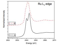
- The above figure shows the Ru L-Edge X-ray absorption spectra for two Ru perovskites; one containing Ru4+ and the other Ru5+. Giving your reasoning, explain which trace corresponds to the Ru4+ oxide and which corresponds to the Ru5+ oxide.
- The two spectra show subtle differences in peak shape around 2860 eV. What is the probable cause of this?
Expert Answer:

Related Book For 
Posted Date:
