

To analyze the sequences in Table 3.2 (see the next page), you will use manual methods that
Question:
Examining the sequences for noticeable differences in length
Comparing the sequences nucleotide by nucleotide
Translating the sequences from codon to amino acid
a. Consider Sequence 1, the oldest sequence from the West Nile region of Egypt, to be the standard for comparison. Examine the sequences shown in the following table for noticeable differences in length. Gaps in sequences are sometimes inserted by the computer as it aligns the rest of the sequence.
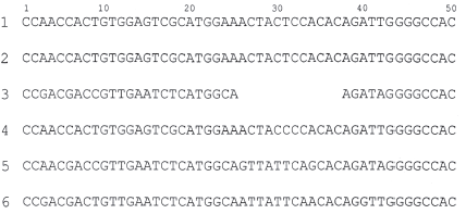
b. Next, analyze the differences in the columns of nucleotides to identify point mutations. Use a straightedge to keep your place, a highlighter, and a pen. Examine each vertical column in above table starting at the left to look for variations from Sequence 1. If the nucleotides in a column match those of the standard sequence, highlight them. If there are deviations from the standard, circle them with the pen. For example, the first column contains all C€™s, so the whole column should be highlighted. The third column has two G€™s, which vary from the A in Sequence 1. The two G€™s would be circled in pen and all the A€™ s highlighted. How many total point mutations did you identify?
c. Determine the percentage of point mutations in sequences 2 through 6 (number of point mutations/ number of nucleotides in sample x 100%). Sequence 2 is done for you as an example. (For Sequence 3, count only the cleotides present in the sequence.)
Sequence 2 = (0/50) x 100% = 0%
Sequence 3 =
Sequence 4 =
Sequence 5 =
Sequence 6 =
Which sample shows the greatest difference in nucleotides from Sequence 1? Explain.
d. In the third and final step in comparing sequences, you need to translate each of the 6 sequences from codon to amino acid, using Figure 17.5 in your textbook. Then you will be able to observe the consequences of the different mutations on the resulting polypeptides. Normally, you would expect to see a start codon (AUG), but assume instead that the reading frame begins with the first nucleotide at the 59 end. Write in the appropriate amino acids under the DNA sequences in above table.
e. Examine each sequence. How many amino acids differed from the standard in sequences 2 through 6? Which amino acids changed?
Sequence 2:
Sequence 3:
Sequence 4:
Sequence 5:
Sequence 6:
What does this information reveal about the effects of the mutations on the E gene and the protein it codes for?
f. How many point mutations were involved in the amino acid differences you found? In above table, draw an asterisk by those nucleotides that made these differences.
g. How many of the point mutations were nonsense mutations? How many were silent mutations?
h. Compare your answers in 2c to those in 2f. Is the percentage of point mutations related to how many amino acids are changed? Explain your response.
i. Is it likely that the deletion mutation is also a frameshift mutation? Explain.
j. Now that you have identified, categorized, and determined the consequences of the various mutations in these sequences of WNV, how do these results compare to your predictions in question 1?
Step by Step Answer:
a These gaps are not present in the actual nucleic acid however they show up in the computers output and often indicate certain kinds of mutations Which of the sequences has either a deletion gaps lea...View the full answer


Campbell Biology
ISBN: 978-0321775658
10th edition
Authors: Jane B. Reece, Lisa A. Urry, Michael L. Cain, Steven A. Wasserman, Peter V. Minorsky, Robert B. Jackson
