

Figure P17.22 shows a microscopic view of muscle tissue. In the figure, structures called sarcomeres are bordered
Question:
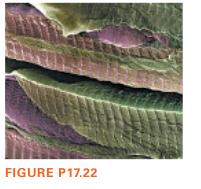
Figure P17.22 shows a microscopic view of muscle tissue. In the figure, structures called sarcomeres are bordered by ridges. The regular pattern of the ridges means that the muscle can operate as a reflection grating, and a measurement of the resulting diffraction pattern can give a measurement of the length of the sarcomeres, which is the distance between the ridges. This has been used to provide measurements of sarcomere length during exercise, as the sarcomeres lengthen and shorten. In one case, an investigator shined a 632 nm laser on exposed muscle tissue in a patient’s forearm as he moved his wrist back and forth through a 100° angle, contracting and then stretching the muscle. The resulting diffraction pattern was projected onto a screen 2.4 cm from the muscle. The investigator measured the distance between the two m = 2 fringes on either side of the central maximum. This length varied from 1.9 cm to 2.7 cm. What were the minimum and maximum values of the sarcomere length?
Step by Step Answer:
To find the minimum and maximum values of the sarcomere length we can use the formula for the distan...View the full answer


College Physics A Strategic Approach
ISBN: 9780134779218
4th Edition
Authors: Randall D. Knight, Brian Jones
