

Question: Consider an MRI scan using a pulse sequence shown in Figure 13.19 in the textbook. A constant x-gradient G_x during each readout time is used,
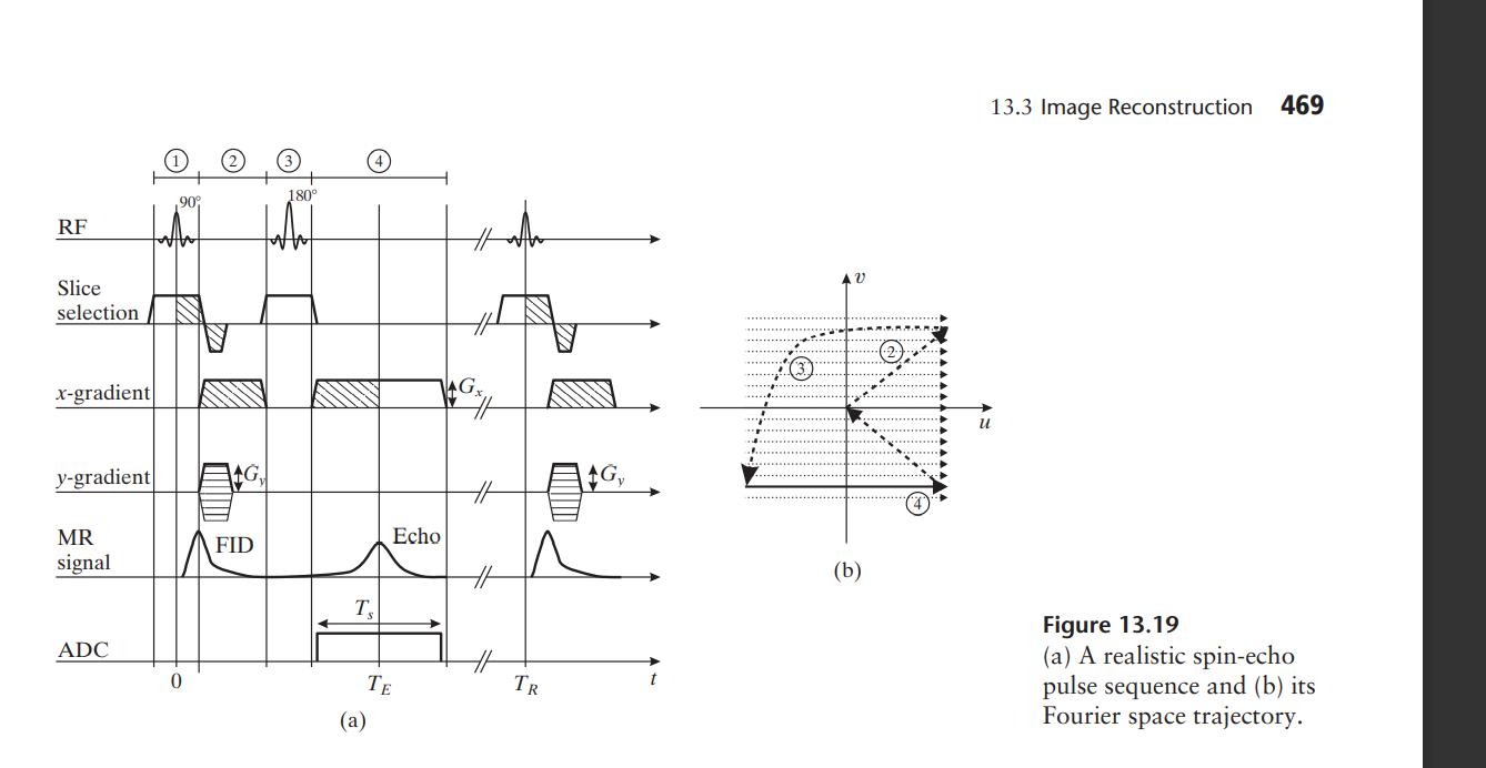 Consider an MRI scan using a pulse sequence shown in Figure 13.19 in the textbook. A constant x-gradient G_x during each readout time is used, and take N samples during each read-out gradient of duration T_s. Also you start with the y-gradient at G_y= deltaG_y M/2, and decrement the y-gradient by deltaG_y each time, and you repeat the process M times. The scan parameters are chosen so that gamma G_xT_s=gamma delta_yT_yM.
(a) What are the sampling patterns in the Frequency domain?
(b) Would the measured signal reflect T2 decay or T2* decay?
(c) If you would like to have a pixel resolution of (d_x,d_y) with d_=d_y=d and covers a rectangular area of FOV_x x FOV_y, with FOV_x= FOV_=W, what are the constraints on the scan parameters? (i.e. give the equations that the parameters G_x,deltaG_y,T_s,T_y, M, N must satisfy in terms of d and W).
(d) What is the reconstructed image dimension?
RF Slice selection x-gradient y-gradient MR signal ADC ,90 G FID 180 T (a) Echo TE TR G AV (b) 13.3 Image Reconstruction 469 u Figure 13.19 (a) A realistic spin-echo pulse sequence and (b) its space trajectory. Fourier
Step by Step Solution
3.41 Rating (164 Votes )
There are 3 Steps involved in it
given Question The pulie Sequence It is the Programmed Set of changing Magnetic gradient ... View full answer

Get step-by-step solutions from verified subject matter experts