New Semester
Started
Get
50% OFF
Study Help!
--h --m --s
Claim Now
Question Answers
Textbooks
Find textbooks, questions and answers
Oops, something went wrong!
Change your search query and then try again
S
Books
FREE
Study Help
Expert Questions
Accounting
General Management
Mathematics
Finance
Organizational Behaviour
Law
Physics
Operating System
Management Leadership
Sociology
Programming
Marketing
Database
Computer Network
Economics
Textbooks Solutions
Accounting
Managerial Accounting
Management Leadership
Cost Accounting
Statistics
Business Law
Corporate Finance
Finance
Economics
Auditing
Tutors
Online Tutors
Find a Tutor
Hire a Tutor
Become a Tutor
AI Tutor
AI Study Planner
NEW
Sell Books
Search
Search
Sign In
Register
study help
sciences
inorganic chemistry
Inorganic Chemistry 5th Edition Catherine Housecroft - Solutions
How many normal modes of vibration are IR active for (a) H2O, (b) SiF4, (c) PCl3, (d) AlCl3, (e) CS2 (f) HCN?
Use the C2v character table to confirm that D2O (‘heavy water’) has three IR active modes of vibration.
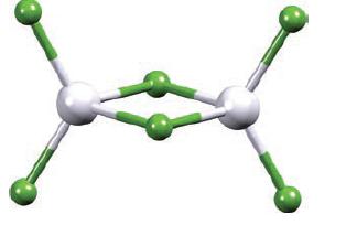
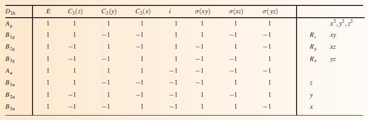
Al2Cl6 belongs to the D2h point group:(a) How many degrees of vibrational freedom does Al2Cl6 possess?(b) Use the D2h character table in Appendix 3 to determine the symmetries of the IR active stretching modes of vibration.Data from Appendix 3 D2h character table.
Six of the nine vibrational degrees of freedom of SiF4 are IR active. Why are IR absorptions observed only at 389 and 1030 cm–1 for this compound?
To what point group does CBr4 belong? Using the appropriate character table, construct a reducible representation for the stretching modes of vibration. Show that this reduces to A1 +T2.
The IR spectra of salts of [AlF6]3– (Oh) exhibit absorptions around 540 and 570 cm–1. Using a group theory approach, confirm that only one of these absorptions arises from a stretching mode.
Determine how many CO stretching modes are possible for trans-M(CO)4X2. What are their symmetries, and how many are IR active?
In 1993, the [Pt(CO)4]2+ ion was reported for the first time [G. Hwang et al. (1993) Inorg. Chem., vol. 32, p. 4667]. One strong absorption at 2235 cm–1 in the IR spectrum was assigned to νCO, and this was absent in the Raman spectrum. In the Raman spectrum, two absorptions (νCO) at 2257 and
Explain how you could distinguish between cis-M(CO)2X2 and trans-M(CO)2X2 by using information from the CO stretching region of IR spectra. Include in your answer a derivation of the number of νCO modes for each molecule.
(a) To which point group does a trigonal bipyramidal XY5 belong? Determine the number and symmetries of the stretching modes of vibration for this molecule.(b) The IR spectrum of gaseous PF5 exhibits absorptions at 1026 and 944 cm–1. Show that this observation is consistent with your answer to
Explain what is meant by the terms (a) Chiral;(b) Enantiomer; (c) Helical chain.
Confirm that the symmetry operation of (a) Inversion is equivalent to an S2 improper rotation, (b) Reflection through a plane is equivalent to an S1 improper rotation.
Open the structure file for problem 3.36: this is the structure of PF5. (a) Orientate the structure so that you are looking down the C3 axis. Where is the σh plane with respect to this axis? (b) Locate three C2 axes in PF5. (c) Locate three σv planes in PF5.(d) To what point group
Haemoglobin is the iron-containing metalloprotein responsible for transporting O2 in the bloodstream of mammals. When O2 binds to the Fe centre, it does so in an ‘end-on’ manner and gives rise to a band in the IR spectrum at 1107 cm−1. No absorption is present in the IR spectrum of gaseous
The conversion of solar energy into chemical energy using artificial photosynthesis involves the photocatalytic conversion of H+ to H2. Why is GC suitable for the detection and quantification of H2?
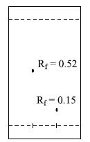
A test TLC plate (silica) using a 1 : 3 mixture of CH3CN:H2O as eluent shows that two compounds have Rf values of 0.52 and 0.15:You plan to use column LC to separate a mixture of the two compounds. How can you attempt to ensure that their behaviour on the column will closely mimic that on the TLC
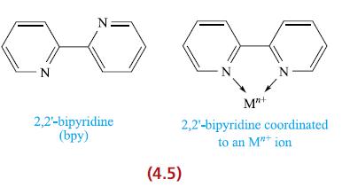
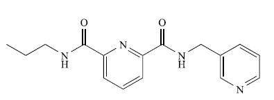
During purification of 2,2'-bipyridine (see structure 4.5), the compound was accidentally exposed to a mineral acid. Elemental analysis gave the following results: C 62.35, H 4.71, N 14.54%. Suggest the identity of the isolated compound.Structure 4.5
What are the near UV and visible ranges in nm? In itz UV-VIS spectrum, Ru3(CO)12 absorbs at 392 nm. Explain why, in a column chromatographic separation of this compound, visual detection is possible.
Why can a CHN analysis of a compound not distinguish between a monomer and dimer of the species? What technique would you use to confirm that a dimer was present?
The reaction of NbCl4(THF)2 with pyridine in the presence of a reducing agent gives NbClx(py)y which contains 50.02% C, 4.20% H and 11.67% N. Determine the values of x and y.
On being heated, the fullerene solvate C60 · xCHBr3 loses solvent in a two-step process. The final weight loss is 41%. Account for these data and determine x.
When gypsum (CaSO4 · 2H2O) is heated to 433 K, it converts to the hemihydrate CaSO4 · 1/2 H2O, and at 463 K, it forms γ-CaSO4. Calculate the % weight changes at 433 and 463 K, and sketch what you expect to see in a TGA curve.
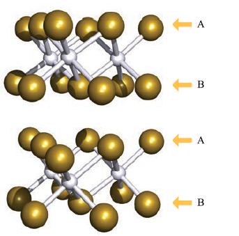
Birnessite, [Na,K][MnIVMnIII]O4 · xH2O, is a mineral with a layered structure of the same type as CdI2 (see Fig. 6.25) comprising octahedral MnO6 units. Na+ and K+ ions and H2O molecules are sited between the layers. The Na/K composition is variable. Analysis for Na is carried out using AAS. A 20
Open the structure file for problem 3.40: this shows the structure of C2Cl6 in the preferred staggered conformation. (a) Orientate the structure so you are looking along the C–C bond. You should be able to see six Cl atoms forming an apparent hexagon around two superimposed C atoms. Why is the
Open the structure file for problem 3.37 which shows the structure of NH2Cl. (a) How many planes of symmetry does NH2Cl possess? (b) Does NH2Cl possess any axes of rotation? (c) Confirm that NH2Cl belongs to the Cs point group. (d) Detail what is meant by the statement: ‘On
Open the structure file for problem 3.38: this shows the structure of OsO4, which has Td symmetry.(a) Orientate the molecule so that you are looking down an O–Os bond, O atom towards you. What rotation axis runs along this bond? (b) The character table for the Td point group shows the
Open the structure file for problem 3.39: this shows the structure of [Co(en)3]3+ where en stands for the bidentate ligand H2NCH2CH2NH2; the H atoms are omitted from the structure. The complex [Co(en)3]3+ is generally described as being octahedral. Look at the character table for the Oh point
Carbon monoxide is a controlled emission from vehicle exhausts. Catalytic converters catalyse the conversion of CO to CO2. Emissions of CO can be quantified using IR spectroscopy with detection limits of <0.5 mgm−3. (a) The IR absorption monitored during analysis for CO is at 2143 cm−1.
Open the structure file for problem 3.41: this shows the structure of α-P4S3. (a) Orientate the structure so that the unique P atom is closest to you and the P3 triangle coincides with the plane of the screen. You are looking down the principal axis of α-P4S3. What type of axis is
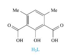
Reaction of H3L (drawn below) with Cu(O2CMe)2 · H2O in MeOH with addition of pyridine (py) yields [Cu4L2(O2CMe)2(py)4(MeOH)2]. Show that a MALDI-TOF mass spectrum with peak envelopes at m/z 977 and 611 is consistent with this formulation. Me ОН ОН H₂L Me ОН
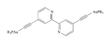
The UV-VIS spectrum of a CH2Cl2 solution of the gold (I) compound shown below with R = Ph is: λmax (ε) = 239 (92500), 269 (67 000), 286 (72 000), 303 (28 000), 315 nm (21000 dm3 mol−1 cm−1).(a) π* ← π transitions contribute to the observed spectrum. How do these arise? (b) Is the
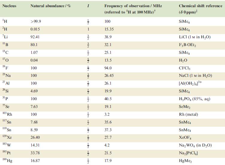
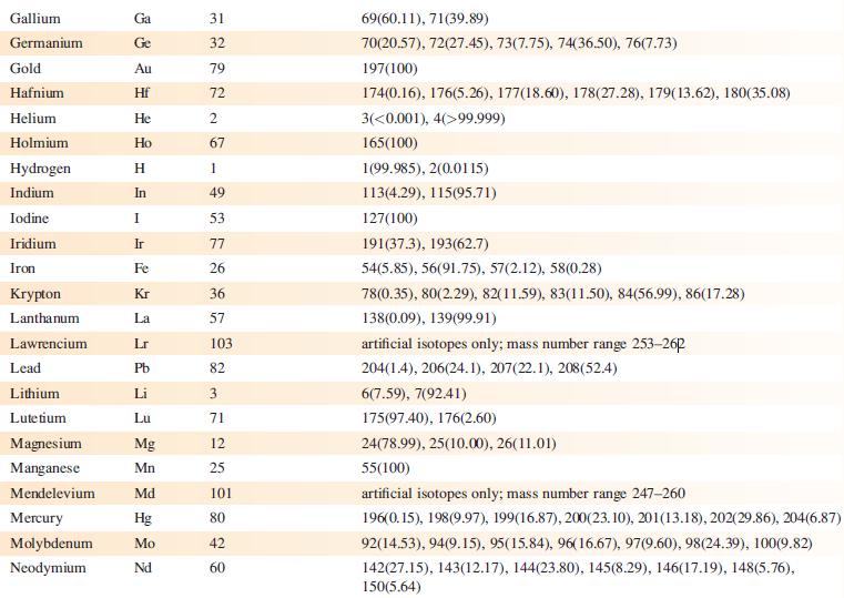
In problems 4.28 to 4.51, refer to Table 4.3 for isotopic abundances where needed.Rationalize the fact that the 13C NMR spectrum of CF3CO2H consists of two binomial quartets with coupling constants of 44 and 284 Hz respectively.Table 4.3 Nucleus ΤΗ ²H Li 11B 13C 170 19 F 23 Na 2 Al 29 Si 31 p 7
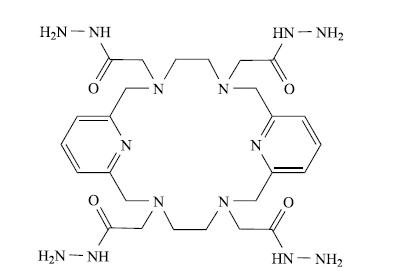
In the MALDI-TOF mass spectrum of the macrocyclic ligand shown below in 1,8,9-trihydroxyanthracene matrix, the dominant peaks are at m/z 615.7 (base peak) and 637.7. Assign the peaks HẠN NH HẠN - NH N N N HN – NH, HN – NH,
The IR spectrum of Li3[PO4] shows absorptions at 1034 and 591 cm−1. There are no bands below the 400 cm−1 cutoff of the IR spectrometer. Why are these data consistent with the [PO4]3− ion being tetrahedral rather than square planar?
The UV-VIS spectrum of a CH3CN solution (2.0 × 10⁻⁵ mol dm−3) of an iron(II) complex is: λmax(ε) = 245 (48 200), 276 (74 100), 284 (81 700), 324 (45 100), 569 nm (25 000 dm3 mol−1 cm−1). A quartz cuvette with path length 1 cm was used for the measurement. (a) Explain why the
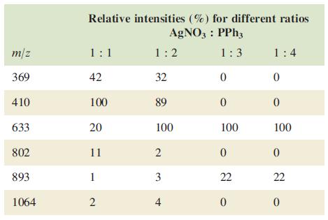
Four MeCN solutions were made up containing AgNO3 and PPh3 in molar ratios of 1 : 1, 1 : 2, 1 : 3 and 1 : 4, respectively. ESI mass spectra (positive mode) of the solutions were recorded, and the data are tabulated below. Account for the data, including the differences between the spectra. Comment
The ESI mass spectrum (positive mode) of the complex shown below contained a peak envelope with m/z 527.9 (100%), 528.9 (15%), 529.9 (46%), 530.9 (7%), 531.9 (0.5%). A group of peaks of low intensity and with spacings of m/z = 1 was also observed around m/z 994. (a) What is the oxidation state of
TheESImass spectrum(positivemode) of the compound shown below exhibits two peaks at m/z 299.2 (base peak) and 321.1. (a) What is a ‘base peak’? (b) Suggest how the observed peaks arise. [Data: C.J. Sumby et al. (2009) Tetrahedron, vol. 65, p. 4681.]Data from Appendix 5 H IZ IN Z O
The EI mass spectrum of lead(II) acetate shows four peak envelopes, each with an isotope pattern characteristic of Pb. The most intense peak in each envelope appears at m/z 326.0, 267.0, 224.0 and 208.0, respectively. (a) By using Appendix 5, sketch the pattern of each peak envelope. (b) Assign
Both positive and negative-ion ESI mass spectra of [Me4Sb][Ph2SbCl4] were recorded. In one spectrum, peaks at m/z 181 (100%), 182 (4.5%), 183 (74.6%) and 184 (3.4%) were observed. The other mass spectrum revealed peaks at m/z 415 (48.8%), 416 (6.4%), 417 (100%), 418 (13.1%), 419 (78.6%), 420
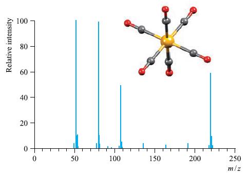
The EI mass spectrum and structure of Cr(CO)6 is shown in Fig. 4.38. Rationalize the peaks in the spectrum. Why is the EI technique suitable for recording the mass spectrum of Cr(CO)6?Figure 4.38 Relative intensity 100 80 80 40 20 T O 50 100 150 200 T 250 m/z
In problems 4.28 to 4.51, refer to Table 4.3 for isotopic abundances where needed.Why is a coupling constant measured in Hz and is not recorded as a chemical shift difference?Table 4.3 Nucleus ΤΗ ²H Li 11B 13C 170 19 F 23 Na 2 Al 29 Si 31 p 7 Se 103 Rh 117. Sn 119 Sn 19 Xe 183 W 195 pt 199
In problems 4.28 to 4.51, refer to Table 4.3 for isotopic abundances where needed.How might you use 31P NMR spectroscopy to distinguish between Ph2PH and Ph3P?Table 4.3 Nucleus ΤΗ ²H Li 11B 13C 170 19 F 23 Na 2 Al 29 Si 31 p 7 Se 103 Rh 117. Sn 119 Sn 19 Xe 183 W 195 pt 199 Hg Natural
In problems 4.28 to 4.51, refer to Table 4.3 for isotopic abundances where needed.Long-range couplings are often observed between 31P and 19F nuclei, between 31P and 1H nuclei, but not between remote non-equivalent 1H nuclei. What does this tell you about the relative magnitudes of values of JPF,
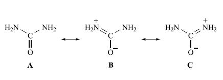
Resonance structures for urea are represented below:The IR spectrum of free urea has absorptions at 3500 and 3350 (ν(NH2)), 1683 (ν(CO)) and 1471 cm−1 (ν(CN)). Urea can bond to metal ions through either an N- or O-donor atom. When urea bonds through the O atom, the contribution from resonance
In the FAB mass spectrum of [Pd(PPh3)4] with NOBA matrix, the base peak appears at m/z 279.1. The isotope pattern shows that Pd is absent from the ion. Suggest an identity for the ion. Why can a peak at m/z 154.0 be ignored?
What is the rule of mutual exclusion? Give two examples of molecular species to which this rule applies.
The ν3 vibrational wavenumber for [BF4]− comes at 1070cm−1, whereas the corresponding band for [BCl4]−, [BBr4]− and [BI4]− comes at 722, 620 and 533 cm−1, respectively. Rationalize this trend.
Vibrational wavenumbers for K[N3] are 2041, 1344 and 645 cm−1. Draw the structure of the [N3]− ion and sketch the three vibrational modes. Which are IR active?
Two isomers, A and B, of a complex can be distinguished because the UV-VIS spectrum of B is blue shifted with respect to that of A. Explain what this means. What is another term used for a blue shift?
Showing 1900 - 2000
of 1950
First
6
7
8
9
10
11
12
13
14
15
16
17
18
19
20
Step by Step Answers